. 2010 Jan 15;165(2):244-54.
doi: 10.1016/j.ygcen.2009.07.006. Epub 2009 Jul 10.

Suppressed production of methyl farnesoid hormones yields developmental defects and lethality in Drosophila larvae

Affiliations

Suppressed production of methyl farnesoid hormones yields developmental defects and lethality in Drosophila larvae

Davy Jones et al. Gen Comp Endocrinol. .

Abstract

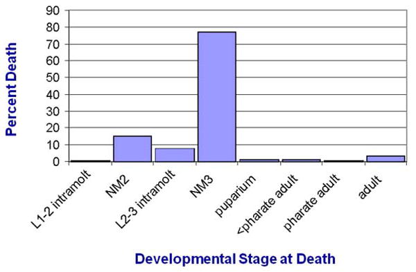
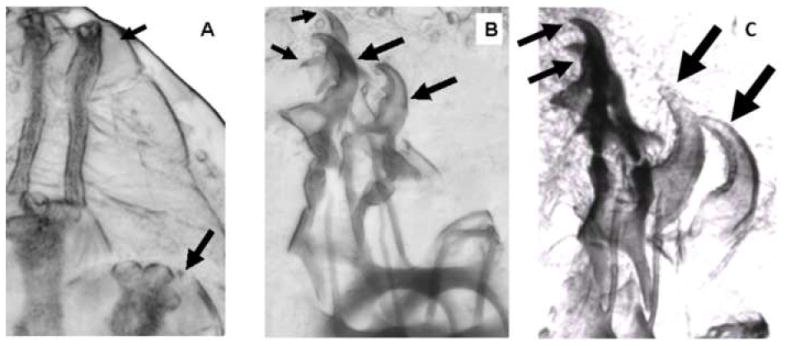
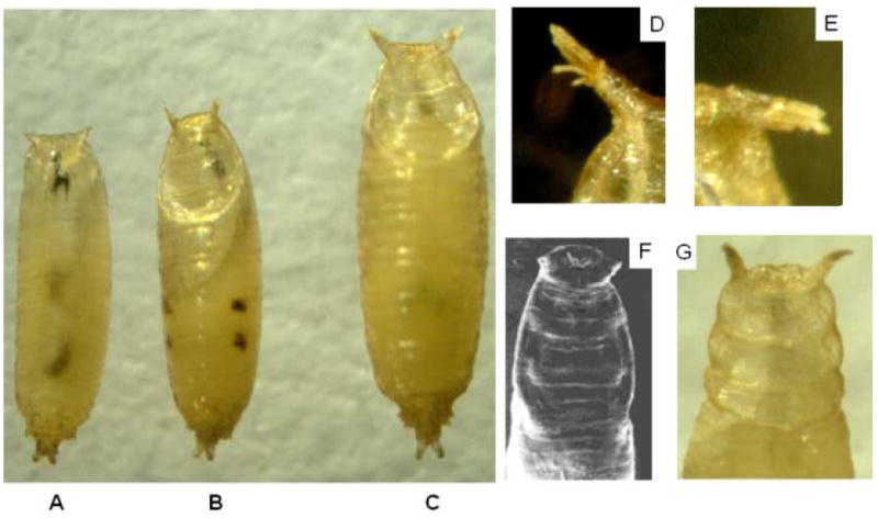
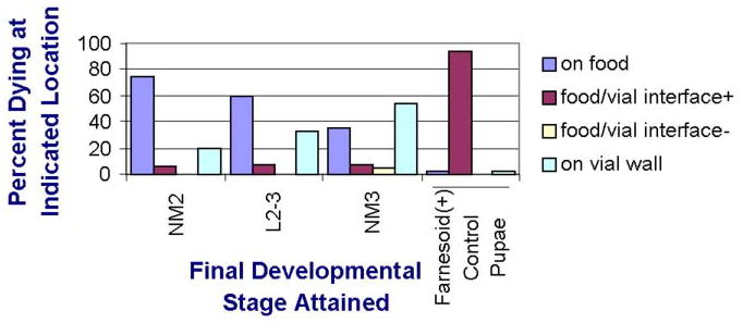
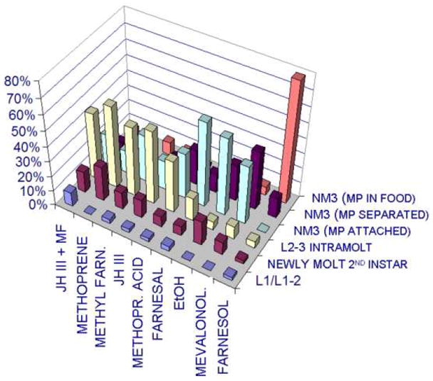
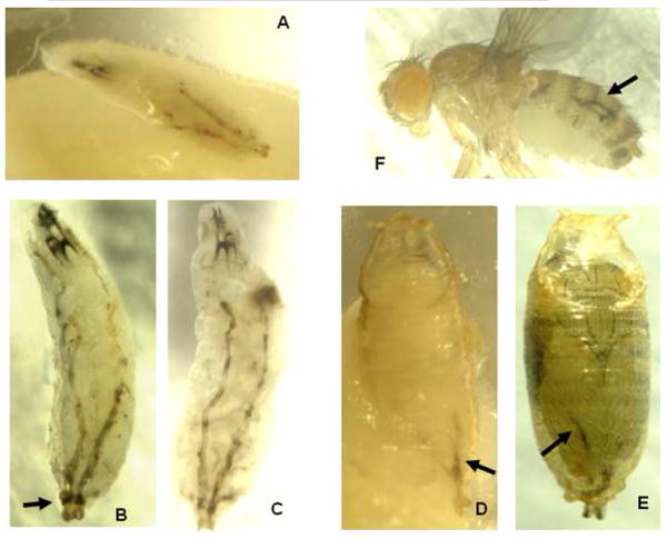
A long-unresolved question in the developmental biology of Drosophila melanogaster has been whether methyl farnesoid hormones secreted by the ring gland are necessary for larval maturation and metamorphosis. In this study, we have used RNAi techniques to inhibit 3-Hydroxy-3-Methylglutaryl CoA Reductase (HMGCR) expression selectively in the corpora allatal cells that produce the circulating farnesoid hormones. The developing larvae manifest a number of developmental, metabolic and morphogenetic derangements. These defects included the exhibition of an "ultraspiracle" death phenotype at the 1st to 2nd instar larval molt, similar to that exhibited by animals that are null for the farnesoid receptor ultraspiracle. The few larvae surviving past a second lethal period at the 2nd to 3rd instar larval molt, again with "ultraspiracle" phenotype, often became developmentally arrested after either attaining a misformed puparium or after formation of the white pupa. Survival past the "ultraspiracle" lethal phenotype could be rescued by dietary provision of an endogenous dedicated precursor to the three naturally secreted methyl farnesoid hormones. In addition to these developmental and morphogenetic defects, most larvae that survived to the late second instar exhibited a posterior-originating melanization of the tracheal system. These results support the hypothesis that larval methyl farnesoid hormones are necessary for larval survival and morphogenetic transformation through the larval and pupal metamorphic processes.


Figures

Figure 1
Figure 1
Mortality of farnesoid (-) larvae at successive developmental stages. Newly hatching 1st instar larvae expressing an RNAi that suppresses expression of HMGCR in the larval corpora allatal cells were moved to individual small diet vials and observed regularly for survival. >99% of these 1st instar larvae burrowed into the diet soon after transfer, and were then subsequently monitored for attainment of the indicated life stages (1-2 intramolt = lst instar larva exhibited incomplete ecdysis to the 2nd instar; NM2 = newly molted 2nd instar; 2-3 intramolt = 2nd instar larva exhibited incomplete ecdysis to the 3rd instar; NM3 = newly molted 3rd instar; puparium = 3rd instar larva attained puparium formation but had not everted to white pupa; < pharate adult = had attained at least white pupa stage but not attained pharate adult status). n = 341
Figure 2
Figure 2
Classic “Ultraspiracle” death phenotype of farnesoid (-) larvae. Some of the larvae expressing an RNAi that suppresses expression of HMGCR in the larval corpora allatal cells were individually reared and observed to developmentally arrest and die after the 1st instar apolysis but prior to ecdysis to the 2nd instar. The view in (A) shows the juxtaposition of the new 2nd instar spiracles (large arrows) to the prior 1st instar spiracles (small arrows). The anterior end view in (B) shows the juxtaposition of the new 2nd instar mouthparts (large arrows) to the prior 1st instar mouthparts (small arrows). The corresponding phenotype was also observed in many larvae that survived to the 2nd to 3rd instar molt. Panel C shows the “double mouthhooks” of 2nd instar larvae (small arrows) and 3rd instar larvae (large arrows).
Figure 3
Figure 3
Failed 2nd to 3rd instar ecdysis phenotype of farnesoid (-) larvae. Some of the larvae expressing an RNAi that suppresses expression of HMGCR in the larval corpora allatal cells were individually reared and were to observed to partially ecdyse but fail to completely ecdyse to the 3rd instar. The most frequently observed phenotypes of these larvae were (Panel A) the splitting of the 2nd instar exoskeleton at the middle of the body but failure to remove the exoskeleton from either of the anterior or posterior ends, (Panel B) the 2nd instar mouthparts remaining caught in the 3rd instar mouthparts, and (Panel C) the posterior end of the 2nd instar exoskeleton remaining caught on the posterior end of the 3rd instar, causing the remainder of the cast 2nd instar exoskeleton (including 2nd instar mouthparts, arrow) to be ‘dragged along.’
Figure 4
Figure 4
Reduced pupa inhibits expression of HMGCR in the larval corpora allatal cells survived to form puparia that then died without pupation. Typically, these larvae were delayed reaching the pupariation stage, and their puparial size (A, ventral view; B, dorsal view) was distinctly less than normal (C). In a frequent phenotype, the projected papillae were appressed together (D, E). Some 3rd instar larvae did not properly contract the body in forming a puparium, especially at the anterior end involving the operculum (G), similar to that reported in Fig. 1 of Henrich et al., (2000), reprinted here as panel F.
Figure 5
Figure 5
Behavioral death phenotype of farnesoid (-) larvae, which exhibited a significantly less incidence of dying at the glass vial/food interface than was the incidence of control animals pupating at that location (P 0 < 0.0001, χ2). Those farnesoid (-) larvae that progressed to L2-3 intramolt before death exhibited a significantly greater incidence of moving up the side of the glass rearing vials just before death than those than died in 2nd instar (P < 0.0001, χ2), and those that attained the newly molted 3rd instar before dying exhibited yet significantly more greater incidence than those dying at L2-3 intramolt, P < 0001, χ2). NM2 = newly molted 2nd instar; L2-3= intramolt from 2nd to 3rd instar; NM3 = newly molted 3rd instar. Interface + = body orientation with head upward onto glass wall and posterior down onto food (see Fig. 10 A). Interface - = body orientation with head downward onto the food and with the posterior upward onto the glass. (n= 400).
Figure 6
Figure 6
Effect (expressed as % death) of provision of farnesoid-like compounds in the larval diet of farnesoid (-) larva. The indicated compounds were layered onto the surface of the larval diet, providing both topical and dietary exposure. Control ethanol-treated larvae did not successfully ecdyse the 2nd instar cuticle, or if they did so, performed the ecdysis on the (abnormal location) above the food surface. However, nearly all larvae exposed to farnesol successfully ecdysed the 2nd instar cuticle to a free 3rd instar, and they performed that ecdysis with the cast 2nd instar mouthparts remaining under the surface of the food, as normal. MF = methyl farnesoate; JH III = methyl epoxyfarnesoate; Methyl Farn. = methyl farnesoate; Methopr. Acid = methoprene acid; Mevalonol. = mevalonolactone; L1 = death during 1st instar; L1-2 = death during 1st instar to 2nd instar intramolt stage prior to ecdysis; L2-3 = death during 2nd instar to 3rd instar intramolt stage prior to ecdysis; NM3 = newly molted 3rd instar; MP attached = 2nd instar mouthparts visible less than one body length away from the dead body of the 3rd instar larva; MP separated = 2nd instar mouthparts visibly located (on food surface or glass vial) more than one body length away from the dead body of the 3rd instar larva; MP in food = 2nd instar mouthparts were not visible on food surface or up on wall of glass vial above food. Dissection of food in several vials confirmed the 2nd instar mouthparts and cuticle were buried under the food surface.
Figure 7
Figure 7
Abnormal melanization of tracheae in farnesoid (-) larvae. Most of the individually reared larvae expressing an RNAi that blocks expression of HMGCR in the corpora allatal cells were to observed exhibit tracheal melanization. The melanization started at the posterior end of the 2 main dorsal tracheal trunks, and rapidly spread anteriorly at or shortly after the 2nd instar to 3rd instar apolysis (A, B). In some cases, there seemed to be a positional periodicity to foci of the most darkened areas (C). Rarely, larvae that exhibited a lesser amount of melanization survived to pupate, and exhibit melanized streaks in the abdomen (D, E), and even more rarely, those pupae would survive to emerge as adults that still exhibited the tracheal melanization (F).

Similar articles

Cited by

References

    1. Barry J, Wang S, Wilson TG. Overexpression of Methoprene-tolerant, a Drosophila melanogaster gene that is critical for juvenile hormone action and insecticide resistance. Insect Biochem Mol Biol. 2008;38:346–53. - PMC - PubMed
    1. Belgacem YH, Martin JR. Hmgcr in the corpus allatum controls sexual dimorphism of locomotor activity and body size via the insulin pathway in Drosophila. PLoS ONE. 2007;2(1):e187. - PMC - PubMed
    1. Bellés X, Martín D, Piulachs MD. The mevalonate pathway and the synthesis of juvenile hormone in insects. Annu Rev Entomol. 2005;50:181–99. - PubMed
    1. Bernardo TJ, Dubrovskaya VA, Jannat H, Maughan B, Dubrovsky EB. Hormonal regulation of the E75 gene in Drosophila: identifying functional regulatory elements through computational and biological analysis. J Mol Biol. 2009;387:794–808. - PubMed
    1. Bialecki M, Shilton A, Fichtenberg C, Segraves WA, Thummel CS. Loss of the ecdysteroid-inducible E75A orphan nuclear receptor uncouples molting from metamorphosis in Drosophila. Dev Cell. 2002;3:209–20. - PubMed

Publication types

MeSH terms

Substances