. 2009 Jul 10;35(1):105-15.
doi: 10.1016/j.molcel.2009.05.026.

Srs2 disassembles Rad51 filaments by a protein-protein interaction triggering ATP turnover and dissociation of Rad51 from DNA

Affiliations

Srs2 disassembles Rad51 filaments by a protein-protein interaction triggering ATP turnover and dissociation of Rad51 from DNA

Edwin Antony et al. Mol Cell. .

Abstract

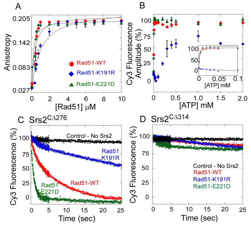
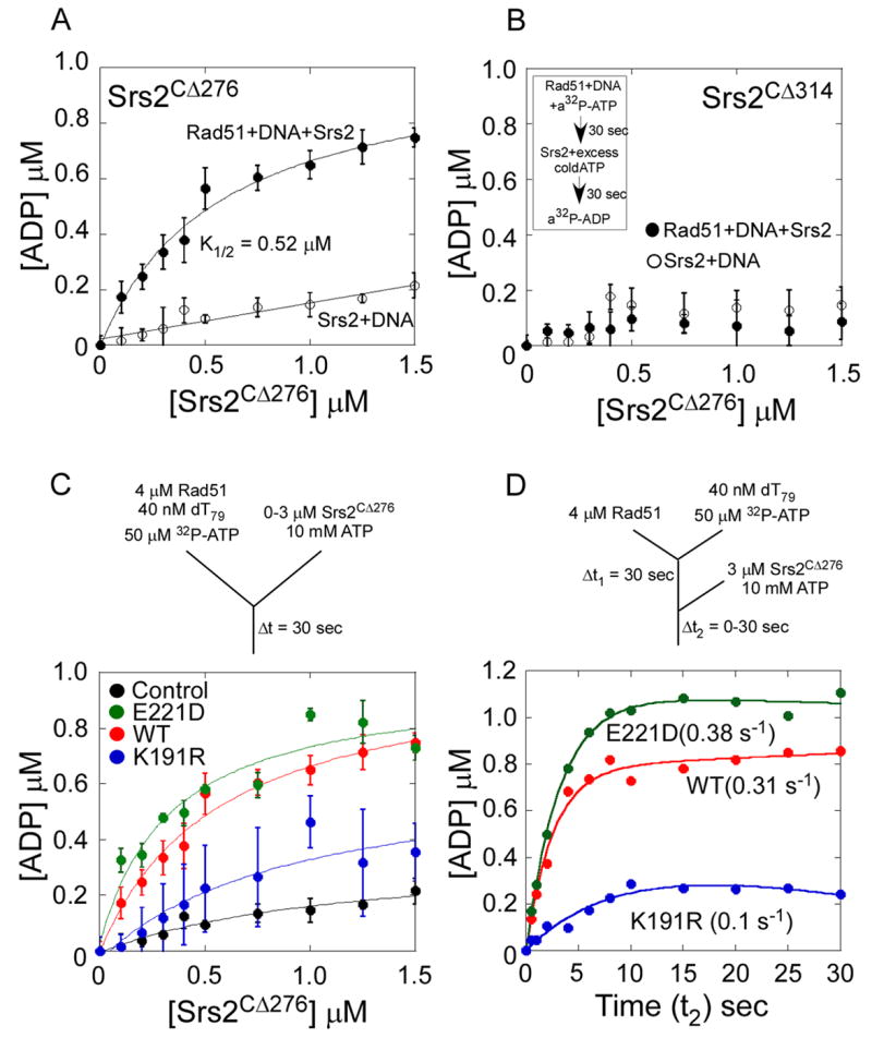
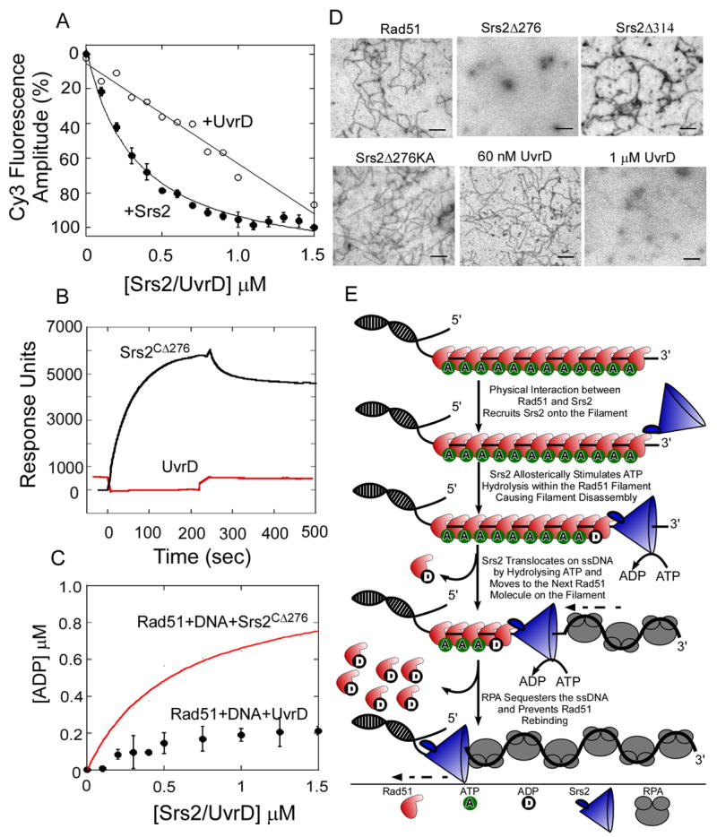
Rad51 is a DNA recombinase functioning in the repair of DNA double-strand breaks and the generation of genetic diversity by homologous recombination (HR). In the presence of ATP, Rad51 self-assembles into an extended polymer on single-stranded DNA to catalyze strand exchange. Inappropriate HR causes genomic instability, and it is normally prevented by remodeling enzymes that antagonize the activities of Rad51 nucleoprotein filaments. In yeast, the Srs2 helicase/translocase suppresses HR by clearing Rad51 polymers from single-stranded DNA. We have examined the mechanism of disassembly of Rad51 nucleoprotein filaments by Srs2 and find that a physical interaction between Rad51 and the C-terminal region of Srs2 triggers ATP hydrolysis within the Rad51 filament, causing Rad51 to dissociate from DNA. This allosteric mechanism explains the biological specialization of Srs2 as a DNA motor protein that antagonizes HR.


Figures

Figure 1
Figure 1. Srs2 Translocates Unidirectionally on ssDNA
(A) Sequence alignment of Saccharomyces cerevisiae Srs2 with various bacterial SF-1 helicases. The conserved helicase motifs, 1A, 2A, 1B and 2B domains in UvrD are depicted. (B) SDS-PAGE analysis of the purified Srs2 proteins used in this study: Srs2CΔ276 and Srs2CΔ314. (C, D) Time courses monitoring Cy3 fluorescence on 5′Cy3 labeled oligonucleotides of varying lengths (25 – 124 nt) shows that Srs2CΔ276 and Srs2CΔ314 translocate unidirectionally on ssDNA.
Figure 2
Figure 2. Assembly and Disassembly of Rad51 Filaments on Single Stranded DNA
(A) Kinetics of Rad51 binding to a 5′Cy3 labeled dT79 oligonucleotide. Increasing concentrations of Rad51 (0-10 μM) were rapidly mixed with 5′Cy3-dT79 ssDNA (40 nM) and ATP-Mg2+ (5 mM) using a stopped flow instrument. The change in Cy3 fluorescence measured as a function of time reveals an increase in the Cy3 fluorescence upon Rad51 binding to the DNA. (B) Kinetics of Rad51 binding to a 3′Cy3 labeled dT79 oligonucleotide. (C) The change in fluorescence amplitude observed in A and B are plotted versus the concentration of Rad51 (log scale). Rad51 binds preferentially to the 5′ end (●) compared to the 3′ end (○) of the ssDNA. (D) Rad51 binding to ssDNA requires nucleotide. Rad51 (4 μM) was rapidly mixed with 5′Cy3-dT79 (40 nM) in the absence (black) or presence of ATP (yellow), ADP (green), AMP-PNP (blue), or ATPγS (red) (5 mM). Change in Cy3 fluorescence was measured over 25 sec post-mixing. (E) The stoichiometry of Rad51 molecules bound to a 5′-FITC labeled dT79 oligonucleotide was measured using fluorescence anisotropy. Increasing concentrations of Rad51 (0–10 μM) were mixed with 5′-FITC labeled dT79 oligonucleotide (40 nM), in the absence (△), or presence of ATP (●), ADP (○), AMP-PNP (▲), or ATPγS (◊) (2.5 mM), and the change in fluorescence anisotropy was measured. (F) Disassociation kinetics of nucleotide-bound Rad51 filaments. Preformed Rad51 filaments −Rad51 (4 μM), 5′Cy3-dT79 (40 nM), and ATP/ADP/AMP-PNP/ATPγS (5 mM), were rapidly mixed with excess unlabeled dT79 (40 μM). Rad51 filaments formed in the presence of ATP (yellow) or AMP-PNP (blue) have longer half-lives: 24.8 s and 55.0 s respectively, compared to ADP (green) or ATPγS bound molecules (red): 0.17 s or 0.09 s respectively. The filaments are stable when not challenged with competitor DNA (black).
Figure 3
Figure 3. Srs2 Clearing of Rad51 Filaments
(A) In stop flow experiments, preformed Rad51 filaments were rapidly mixed with Srs2CΔ276. In the absence of Srs2, Rad51 filaments are stable (blue trace), but when challenged with Srs2 (1.5 μM post-mixing) Rad51 filaments are cleared (red trace). (B) Srs2CΔ276 concentration dependence of Rad51 clearing was measured with increasing concentrations of Srs2CΔ276 (0 - 1.5 μM post-mixing, shown as insert). The relative change in fluorescence amplitude is plotted as a function of Srs2 concentration and yields a K1/2 = 0.38 ± 0.04 μM for Rad51 filament clearing by Srs2CΔ276. (C) The Rad51 filament clearing experiment in A was repeated with 5′Cy3 ssDNA of varying lengths (dTn, n = 59-124 nt). Rad51 (2 μM post-mixing) was used in all the experiments to maintain sub-saturating Rad51-DNA filament forming conditions. The clearing rate observed for each DNA length in the presence of Srs2 (1.5 μM post-mixing) is plotted as a function of DNA length. The red dotted line is the expected trend for the clearing rate if a continuous Rad51-DNA filament is formed under these sub-saturating Rad51 concentrations. (D) The Rad51-DNA filament clearing experiment described in C was repeated with saturating concentrations of Rad51 (10 μM post-mixing). The clearing rates observed under these conditions are plotted as a function of DNA length. The red dotted line represents the expected trend for the clearing rate if Srs2 initiates clearing only at the 3′ end of the Rad51-DNA filament. Deviation from the predicted trend suggests that Srs2 can invade a Rad51 filament at multiple points and initiate clearing. (E) and (F) depict schematic representations of expected Rad51-DNA filaments formed under limiting or saturating concentrations of Rad51, and potential Srs2 loading or invading sites on the respective filaments.
Figure 4
Figure 4. The C-terminus of Srs2 is Essential for its Physical Interaction with Rad51 and Filament Clearing Activity
(A) Pre-formed Rad51-DNA-ATP filaments were rapidly mixed with buffer (blue trace), Srs2CΔ276 (red trace), or Srs2CΔ314 (green trace), and filament clearing monitored as a function of time. Srs2CΔ314, lacking an additional 38 residues is unable to clear a Rad51-DNA filament. (B) Srs2 concentration dependence of filament clearing shows a weak filament clearing activity for Srs2CΔ314 (◆) compared to Srs2CΔ276 (●). (C) An ATP binding/hydrolysis deficient mutant of Srs2CΔ276, Srs2CΔ276-K41A (◆), is unable to clear a preformed Rad51-DNA filament unlike the wild-type Srs2CΔ276 protein (●). (D) A synthetic peptide corresponding to the Srs2 C-terminal residues 861 – 898, is a poor competitor for Srs2CΔ276 in Rad51 filament clearing experiments. Rad51 was tethered onto a SPR chip, and increasing concentrations of (E) Srs2CΔ276, or (F) Srs2CΔ314 was passed over the chip. The insert shows the change in amplitude (response units, RU) plotted versus the concentration of Srs2, yielding a KD = 15.2 ± 5 μM for the interaction between Srs2CΔ276 and Rad51. The interaction between Srs2CΔ314 and Rad51 is very weak.
Figure 5
Figure 5. Rad51 Filament Disassembly is Regulated by ATP Hydrolysis Within the Rad51-DNA Filament
(A) Rad51-WT (●), Rad51-K191R (◆), and Rad51-E221D (▲) proteins form a filament on ssDNA measured by fluorescence anisotropy. The ATP hydrolysis deficient mutant (Rad51-K191R) binds ssDNA with lower affinity (KD = 1.21 ± 0.3 μM) compared to the wild-type Rad51 (KD = 0.28 ± 0.1 μM) or the Rad51-E221D mutant (KD = 0.21 ± 0.1 μM). (B) ATP concentration dependence of Rad51 filament formation on ssDNA measured using the stopped flow assay and monitoring changes in 5′Cy3 fluorescence on a dT79 DNA substrate. Change in fluorescence amplitude observed at each ATP concentration is plotted for wild-type Rad51 (●), Rad51-K191R (◆), and Rad51-E221D (▲). The K1/2 for ATP dependence of filament formation for wild-type Rad51 and Rad51-K191R are 1.5 ± 0.3 nM and 2.1 ± 0.6 nM respectively. The Rad51-K191R mutant (◆) does not bind to DNA at lower ATP concentrations (insert), but forms a filament at higher ATP concentrations (K1/2 = 120 ± 28 nM). (C) Srs2CΔ276 clears wild-type Rad51 (red), Rad51-K191R (blue), and Rad51-E221D (green) filaments, but (D) Srs2CΔ314 is incapable of clearing either wild-type Rad51 or mutant Rad51 filaments.
Figure 6
Figure 6. Srs2 Allosterically Stimulates ATP Hydrolysis within the Rad51 Filament Thereby Promoting Filament Disassembly
(A) Srs2CΔ276 stimulates the ATP hydrolysis within a Rad51-DNA filament. Preformed Rad51-DNA-32P-ATP filaments were mixed with increasing concentrations of Srs2CΔ276 and excess unlabeled ATP and the amount of 32P-ADP formed was measured. A stimulation of ATP hydrolysis is observed within the Rad51 filament (●), and the Srs2CΔ276 dependence of this stimulation yields a K1/2 = 0.53 ± 0.13 μM. The control experiment done in the absence of Rad51 (○) shows the background level of 32P-ATP hydrolyzed by Srs2 when a 500-fold excess of unlabeled ATP is present. (B) Srs2CΔ314 does not stimulate ATP hydrolysis within a Rad51-DNA filament. The insert shows a schematic representation of the ATPase assays described in A and B. (C) Rad51 filaments were formed on ssDNA (dT79) with either wild-type Rad51 (red), Rad51-K191R (blue), or Rad51-E221D (green) proteins in the presence of radiolabeled ATP. Srs2CΔ276 stimulates the rate of ATP hydrolysis when added to these filaments along with excess unlabeled ATP. The Srs2CΔ276 dependence of this stimulation yields a K1/2 = 0.57 ± 0.15 μM (WT), K1/2 = 0.9 ± 0.1 μM (K191R), and K1/2 = 0.34 ± 0.07 μM (E221D) respectively. The control experiment done in the absence of Rad51 (black) shows the background level of 32P-ATP hydrolyzed by Srs2 when a 500-fold excess of unlabeled ATP is present. (D) Rate of ATP hydrolysis within the various Rad51 nucleoprotein filaments during filament clearing by Srs2CΔ276 measured using a sequential mixing quenched flow setup. Rad51 (4 μM, WT-red, K191R-blue or E221D-green) was first mixed with dT79 DNA (40 nM) and 32P-ATP (50 μM) for 30 seconds and then rapidly mixed with Srs2 (3 μM) and 20 mM excess ATP for varying times (t2). The schematic for the sequential mixing experiment is shown, and the Srs2 stimulated rates of ATP hydrolysis in the wild-type and mutant Rad51 filaments are noted.
Figure 7
Figure 7. Mechanism of Clearing Rad51 Filaments by Srs2
(A) In stop flow experiments, preformed Rad51 filaments were rapidly mixed with increasing concentrations of Srs2CΔ276 (●) or UvrD (○). The relative change in fluorescence amplitude is plotted as a function of Srs2/UvrD concentration. (B) Rad51 was tethered onto a SPR chip and 7 μM UvrD (red) or Srs2CΔ276 (black) was passed over the chip. The change in response units (RU) is plotted versus time. Srs2CΔ276 binds to Rad51 but no interaction between Rad51 and UvrD is observed. (C) UvrD does not stimulate the rate of ATP hydrolysis within a Rad51-DNA filament (●) unlike Srs2CΔ276 (red trace). (D) EM analysis of Rad51 filaments formed on M13 mp18(+) ssDNA and challenged with RPA, along with 60 nM Srs2CΔ276 , Srs2CΔ314 , Srs2 Srs2CΔ276 KA, UvrD, or 1 μM UvrD. 60 nM Srs2CΔ276 or 1 μM UvrD are capable of clearing filaments, whereas Rad51 filaments are intact when challenged with 60 nM Srs2CΔ314, Srs2CΔ276 KA, or UvrD. (E) The physical interaction between Srs2 and Rad51 is required for Srs2 to gain access onto the Rad51 nucleoprotein filament. ATP hydrolysis within the Rad51-DNA filament, allosterically modulated by Srs2, promotes filament disassembly, and the ssDNA translocation activity of Srs2 enables processive 3′ to 5′ clearing of the filament. The free DNA is sequestered by RPA and prevents reformation of the Rad51 filament.

References

    1. Bianco PR, Tracy RB, Kowalczykowski SC. DNA strand exchange proteins: a biochemical and physical comparison. Front Biosci. 1998;3:D570–603. - PubMed
    1. Bugreev DV, Yu X, Egelman EH, Mazin AV. Novel pro- and anti-recombination activities of the Bloom’s syndrome helicase. Genes Dev. 2007;21:3085–3094. - PMC - PubMed
    1. Chi P, Van Komen S, Sehorn MG, Sigurdsson S, Sung P. Roles of ATP binding and ATP hydrolysis in human Rad51 recombinase function. DNA Repair (Amst) 2006;5:381–391. - PubMed
    1. Chiolo I, Carotenuto W, Maffioletti G, Petrini JH, Foiani M, Liberi G. Srs2 and Sgs1 DNA helicases associate with Mre11 in different subcomplexes following checkpoint activation and CDK1-mediated Srs2 phosphorylation. Mol Cell Biol. 2005;25:5738–5751. - PMC - PubMed
    1. Conway AB, Lynch TW, Zhang Y, Fortin GS, Fung CW, Symington LS, Rice PA. Crystal structure of a Rad51 filament. Nat Struct Mol Biol. 2004;11:791–796. - PubMed

Publication types

MeSH terms