. 2009 Oct;145(1):166-70.
doi: 10.1016/j.virusres.2009.07.004. Epub 2009 Jul 10.

Participation of the phosphatidylinositol 3-kinase/Akt pathway in Junín virus replication in vitro

Affiliations

Participation of the phosphatidylinositol 3-kinase/Akt pathway in Junín virus replication in vitro

F N Linero et al. Virus Res. 2009 Oct.

Abstract

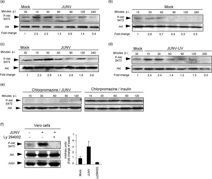
In this paper we demonstrate that infection of cell cultures with the arenavirus Junín (JUNV), agent of the argentine haemorrhagic fever, leads to the activation of PI3K/Akt signalling pathway. Phosphorylation of Akt occurs early during JUNV infection of Vero cells and is blocked by the PI3K inhibitor, Ly294002. Infection of cells with UV-irradiated JUNV redeemed the pattern of stimulation observed for infectious virus indicating that an early stage of multiplication cycle would be enough to trigger activation. Treatment of cells with chlorpromazine abrogated phosphorylation of Akt upon JUNV infection suggesting virus internalization as responsible for activation. Inhibition of Akt phosphorylation by Ly294002 impaired viral protein synthesis and expression leading to a reduced infectious virus yield without blocking the onset of persistent stage of infection. This impairment is linked to a reduced amount of virus bound to cells probably due to a blockage on the recycling of transferrin cell-receptor, employed by the virus to adsorb to the cell surface. Early Akt activation was also observed in BHK-21 and A549 JUNV infected cells suggesting an important role of PI3K/Akt signalling in JUNV multiplication in vitro.


Figures

Fig. 1
Fig. 1
JUNV infection induces Akt phosphorylation via a PI3K-dependent pathway. Vero (a) and BHK-21 (c) cells were infected with XJCl3 strain of JUNV at an MOI of 1 PFU/cell and processed for WB at the indicated times p.i. Mock infected Vero (b and first lines in a and d) and BHK-21 (first line in c) cells and Vero cells infected with UV-inactivated JUNV (d) were used as controls. Vero cells pre-treated for 2 h with 40 μM chlorpromazine were infected with JUNV at an MOI of 1 PFU/cell (e, left panel) or incubated with 240 μM insulin (e, right panel) and processed for WB at indicated times p.i. Vero cells treated with 10 μM Ly294002 (f) were infected with JUNV at an MOI of 1 PFU/cell and processed for WB at 30 min p.i. Phosphorylation of Akt (Ser473) was determined by WB in whole-cell lysates using a phospho-Akt (Ser473) (CST 9271, New England Biolabs) or total Akt (CST 9272, New England Biolabs) antibodies. Fold change of Akt phosphorylation was expressed as densitometric units (Scion Image software) of band normalized to the total Akt level relative to the uninfected control from of each panel. All experiments were performed on 24 h serum starved cells, and the infection and treatments were carried out in serum free medium.
Fig. 2
Fig. 2
Inactivation of PI3K/Akt pathway impairs JUNV multiplication. (a) Vero and BHK-21 cells were infected with JUNV at an MOI of 1  PFU/cell and treated with 10 μM Ly294002 (Sigma, MO). At 24 h p.i. supernatants were collected and virus titer was calculated by plaque assay. Alternatively, cells treated under the same conditions mentioned above were fixed with methanol and subjected to IFA (b) or processed for WB (c) to determine N synthesis and expression. Values in b) indicate % of inhibition calculated from amounts of fluorescence intensities (ImagePro-Express software) in treated cells in comparison to untreated controls. Similar numbers of total cells were analyzed for each category. Vero cells transfected either with pCEFL vector encoding wild-type Akt (PKB-wt) or a kinase-inactive mutant (PKB-kd) were infected with JUNV at an MOI of 10 PFU/cell at 24 h post-transfection and fixed at 24 h p.i. with methanol for IFA, employing anti-N (red fluorescence) and anti-Akt (green fluorescence) antibodies. Values in merge panels indicate % of cells bearing both colors (approx. 150 cells bearing similar levels of Akt expression were scored in duplicate coverslips). After 24 h of infection, virus yield was determined by plaque assay, vesicular stomatitis virus (VSV) was employed as a negative control (e). Vero cells infected with JUNV at an MOI of 1 PFU/cell were treated at different times p.i. with 10 μM Ly294002, further incubated up to 24 h p.i. and virus yield quantified by plaque assay (f). Vero cells were infected with JUNV at an MOI of 10 PFU/cell in the presence or absence of 10 μM Ly294002, incubated at 37 °C and lysed by freeze and thawing at different times post-inoculation (g). Internalized virus in cell lysates was quantified by plaque assay. Vero cells treated or not with 10 μM Ly294002 were incubated with TRITC-labeled transferrin (SIGMA) and processed for IFA at 15 and 30 min post-contact (h). Vero cells previously incubated with 20 μg/ml transferrin (Tf) or 10 μM Ly294002 (Ly) or both transferrin and Ly294002 (Ly + Tf) were infected with approx. 150 PFU of JUNV and overlayed with plaquing medium (i). All experiments (n = 3) were performed on 24 h serum starved cultures and infection and/or treatment were carried out in serum free medium.

References

    1. Bowick G.C., Fennewald S.M., Scott E.P., Zhang L., Elsom B.L., Aronson J.F., Spratt H.M., Luxon B.A., Gorenstein D.G., Herzog N.K. Identification of differentially activated cell-signaling networks associated with Pichinde virus pathogenesis by using systems kinomics. J. Virol. 2007;81:1923–1933. - PMC - PubMed
    1. Briand G., Barbeau B., Tremblay M. Binding of HIV-1 to its receptor induces tyrosine phosphorylation of several CD4-associated proteins, including the phosphatidylinositol 3-kinase. Virology. 1997;228:171–179. - PubMed
    1. Buchkovich N.J., Yu Y., Zampieri C.A., Alwine J.C. The TORrid affairs of viruses: effects of mammalian DNA viruses on the PI3K-Akt-mTOR signalling pathway. Nat. Rev. Microbiol. 2008;6:266–275. - PMC - PubMed
    1. Buchmeier M.J. Arenaviruses: protein structure and function. Curr. Top. Microbiol. Immunol. 2002;262:159–173. - PubMed
    1. Cooray S. The pivotal role of phosphatidylinositol 3-kinase-Akt signal transduction in virus survival. J. Gen. Virol. 2004;85:1065–1076. - PubMed

Publication types

MeSH terms

Substances

LinkOut - more resources