Review
. 2009 Sep-Oct;1789(9-10):667-74.
doi: 10.1016/j.bbagrm.2009.06.005. Epub 2009 Jul 9.

The toll-like receptor 3:dsRNA signaling complex

Affiliations
Review

The toll-like receptor 3:dsRNA signaling complex

Istvan Botos et al. Biochim Biophys Acta. 2009 Sep-Oct.

Abstract

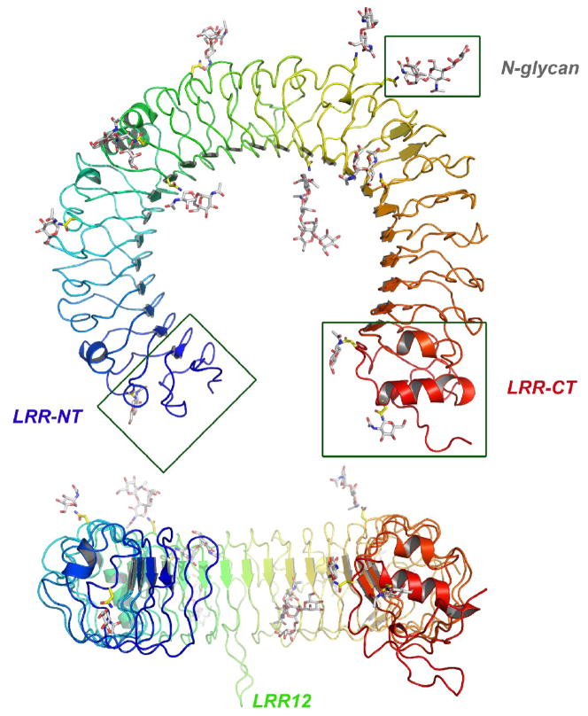
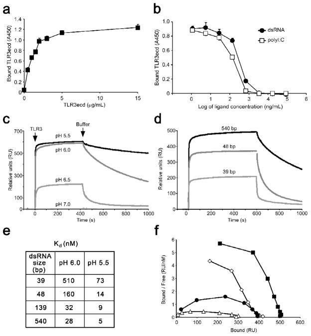
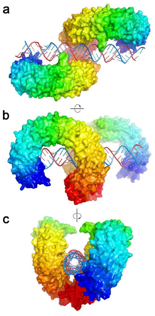
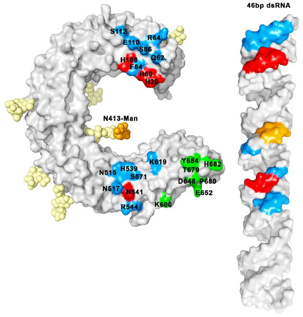
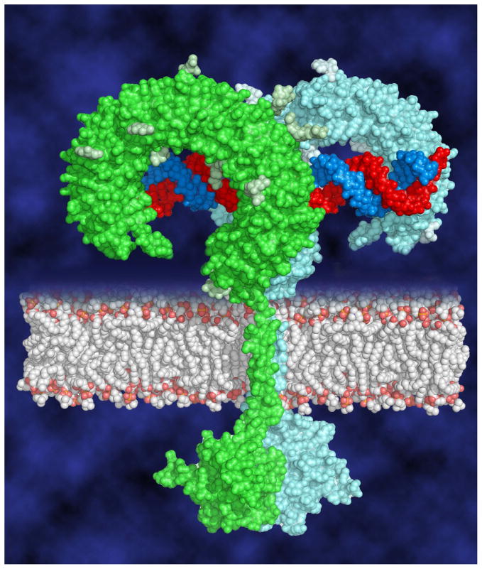
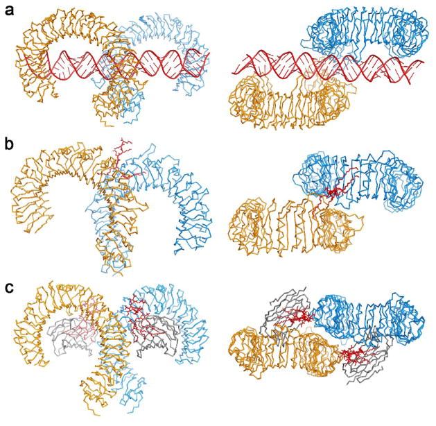
Toll-like receptors (TLRs) recognize conserved molecular patterns in invading pathogens and trigger innate immune responses. TLR3 recognizes dsRNA, a molecular signature of most viruses via its ectodomain (ECD). The TLR3-ECD structure consists of a 23 turn coil bent into the shape of a horseshoe with specialized domains capping the N and C-terminal ends of the coil. TLR3-ECDs bind as dimeric units to dsRNA oligonucleotides of at least 45 bp in length, the minimal length required for signal transduction. X-ray analysis has shown that each TLR3-ECD of a dimer binds dsRNA at two sites located at opposite ends of the TLR3 "horseshoe" on the one lateral face that lacks N-linked glycans. Intermolecular contacts between the C-terminal domains of two TLR3-ECDs stabilize the dimer and position the C-terminal residues within 20-25 A of each other, which is thought to be essential for transducing a signal across the plasma membrane in intact TLR3 molecules. Interestingly, in TLRs 1, 2 and 4, which bind lipid ligands using very different interactions from TLR3, the ligands nevertheless promote the formation of a dimer in which the same two lateral surfaces as in the TLR3-ECD:dsRNA complex face each other, bringing their C-termini in close proximity. Thus, a pattern is emerging in which pathogen-derived substances bind to TLR-ECDs, thereby promoting the formation of a dimer in which the glycan-free ligand binding surfaces face each other and the two C-termini are brought in close proximity for signal transduction.


Figures

Fig. 1
Fig. 1
Structure of the TLR3-ECD. The TLR3-ECD consists of 23 LRRs that form a horseshoe-like solenoid with two capping motifs. The molecule has a surprisingly flat profile. There are 11 visible glycosylation sites.
Fig. 2
Fig. 2
The interaction of soluble TLR3-ECD with dsRNA. (a,b) ELISA showing the binding of TLR3-ECD to immobilized 540 bp dsRNA. (a) binding at increasing TLR3-ECD concentrations, (b) inhibition of binding by free 540 bp dsRNA or polyI:C. (c) Binding and dissociation of TLR3-ECD to immobilized 540 bp dsRNA at indicated pH values, using surface plasmon resonance (SPR). The TLR3-ECD was added at the arrow labeled TLR3, and was replaced by medium at the arrow Buffer. (d) TLR3-ECD binding at pH 6.0 to increasing lengths of immobilized dsRNA measured by SPR. (e) Dissociation constants of TLR3-ECD binding to varying lengths of dsRNA at pH 6.0 and 5.5, calculated from SPR data. (f) Scatchard plot of binding data at pH 6.0. Downward curvature indicates positive cooperativity. Data are taken from [18].
Fig. 3
Fig. 3
Structure of the TLR3-ECD:dsRNA signaling complex. (a–c) 3 views of the TLR3-ECD dimer bound to a 46-mer dsRNA. (a,b) The glycan-free face of each TLR3 interacts with the dsRNA. There are two binding sites for each TLR3 molecule: C-terminal and N-terminal. (c) Looking down the axis of the dsRNA shows that dsRNA is linear and the C-termini of TLR3-ECD interact. The N-terminal region is colored purple, and the C-terminal, red.
Fig. 4
Fig. 4
Residues involved in binding. Essential residues (which lead to loss of function when mutated to alanine) (in red) and interacting non-essential residues (which make contact with dsRNA in the crystal structure but retain function when mutated)(in blue) are mapped on the surface of the ECD with the corresponding colors on the dsRNA. Carbohydrate-ligand interactions are highlighted in orange. Some residues in the 600–684 range (in green) are involved in the homotypic protein-protein interactions between the two TLR-ECDs. The N-terminal ligand binding site is formed by labeled residues from the 39–112 range, and the C-terminal site by labeled residues from 515–619.
Fig. 5
Fig. 5
Model of the full-length signaling complex. Two TLR3 ECDs associate on the dsRNA ligand bringing the cytoplasmic TIR domains together to initiate downstream signaling.
Fig. 6
Fig. 6
Crystal structures of TLR signaling complexes. (a) side and top views of the TLR3:dsRNA homodimer [19] (b) TLR1/TLR2:Pam3CSK4 heterodimer [12] and (c) TLR4/MD2:LPS homodimer [21]. The ligands are shown in red.

Similar articles

Cited by

References

    1. Bell JK, Askins J, Hall PR, Davies DR, Segal DM. The dsRNA binding site of human Toll-like receptor 3. Proc Natl Acad Sci U S A. 2006;103:8792. - PMC - PubMed
    1. Bell JK, Botos I, Hall PR, Askins J, Shiloach J, Segal DM, Davies DR. The molecular structure of the Toll-like receptor 3 ligand-binding domain. Proc Natl Acad Sci U S A. 2005;102:10976. - PMC - PubMed
    1. Bell JK, Mullen GE, Leifer CA, Mazzoni A, Davies DR, Segal DM. Leucine-rich repeats and pathogen recognition in Toll-like receptors. Trends Immunol. 2003;24:528. - PubMed
    1. Choe J, Kelker MS, Wilson IA. Crystal structure of human toll-like receptor 3 (TLR3) ectodomain. Science. 2005;309:581. - PubMed
    1. de Bouteiller O, Merck E, Hasan UA, Hubac S, Benguigui B, Trinchieri G, Bates EE, Caux C. Recognition of double-stranded RNA by human toll-like receptor 3 and downstream receptor signaling requires multimerization and an acidic pH. J Biol Chem. 2005;280:38133. - PubMed

LinkOut - more resources