. 2009 Oct 30;184(1):142-51.
doi: 10.1016/j.jneumeth.2009.07.005. Epub 2009 Jul 21.

Investigating the depth electrode-brain interface in deep brain stimulation using finite element models with graded complexity in structure and solution

Affiliations

Investigating the depth electrode-brain interface in deep brain stimulation using finite element models with graded complexity in structure and solution

Nada Yousif et al. J Neurosci Methods. .

Abstract

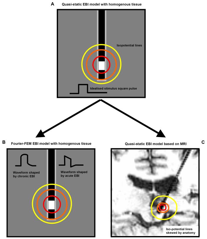
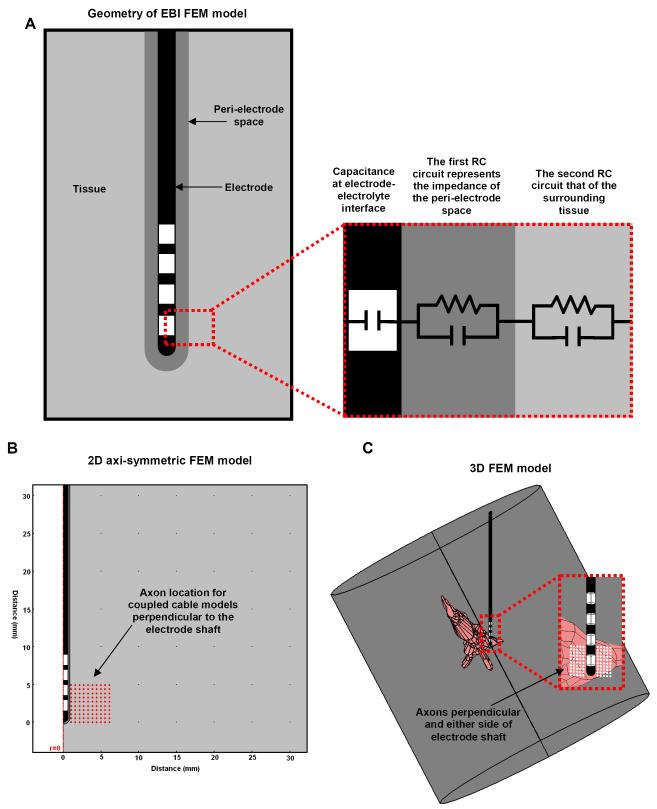
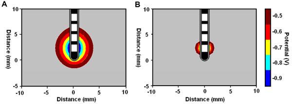
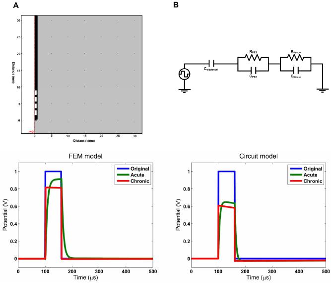
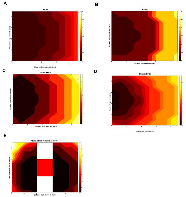
Deep brain stimulation (DBS) is an increasingly used surgical therapy for a range of neurological disorders involving the long-term electrical stimulation of various regions of the human brain in a disorder specific manner. Despite being used for the last 20 years, the underlying mechanisms are still not known, and disputed. In particular, when the electrodes are implanted into the human brain, an interface is created with changing biophysical properties which may impact on stimulation. We previously defined the electrode-brain interface (EBI) as consisting of three structural elements: the quadripolar DBS electrode, the peri-electrode space and the surrounding brain tissue. In order to understand more about the nature of this EBI, we used structural computational models of this interface, and estimated the effects of stimulation using coupled axon models. These finite element models differ in complexity, each highlighting a different feature of the EBI's effect on the DBS-induced electric field. We show that the quasi-static models are sufficient to demonstrate the difference between the acute and chronic clinical stages post-implantation. However, the frequency-dependent models are necessary as the waveform shaping has a major influence on the activation of neuronal fibres. We also investigate anatomical effects on the electric field, by taking specific account of the ventricular system in the human brain. Taken together, these models allow us to visualise the static, dynamic and target specific properties of the DBS-induced field in the surrounding brain regions.


Figures

Figure 1
Figure 1
The process of developing the model of the EBI in the FEM formulism is schematised here. The starting point is the simplistic quasi-static model (A). This assumes that the solution has no frequency or time dependence. This can be extended into a Fourier FEM model to allow for the frequency dependence of tissue, and therefore represent the waveform shaping over time (B). The geometry of the model can also account for the anatomical features of the implantation site as obtained from a post-operative MRI scan (C).
Figure 2
Figure 2
(A) Schematic representation of the electrode-brain interface (EBI). We defined the EBI as consisting of the DBS electrode, the surrounding tissue, and a peri-electrode space whose properties change over time. The EBI can also be represented by an equivalent circuit (A right). (B) This can be modelled using a 2-dimensional axi-symmetric representation as this geometry is symmetrical along the axis of the electrode, which has the advantage of using less computational power to simulate. (C) However, a 3-dimensional model represents the precise geometry of the electrode, located within a cylinder of surrounding tissue which is centred on the tip of the electrode but orientated along the axis of the anatomical details included, in this case the third ventricle and cerebral aqueduct. Such FEM models were combined with axon models, which can be orientated perpendicular to both the electrode shaft, and the plane of the axi-symmetric model (red/white circles).
Figure 3
Figure 3
Quasi-static model solutions showing the effects of the changing biophysics of the EBI induced in the acute (A) and the chronic (B) stages post-implantation on the induced potential distribution with a stimulus amplitude of -1V.
Figure 4
Figure 4
Both the Fourier-FEM model and the equivalent circuit model reveal the frequency dependent effects of the interface on the stimulus waveform. The circuit model represents the complex impedance of the peri-electrode space and the tissue using a RC circuit for each compartment. In the FEM model, these properties are represented by the conductivity and the permittivity of the regions in the geometry. The electrode is assumed to be perfectly polarisable and is consequently modelled as a pure capacitance. In both cases, the acute interface demonstrates low-pass filtering behaviour, whereas in the chronic stage the effect is mainly a reduction in amplitude. The charge delivered can be measured by the area under the curve, and the FEM model results show that 93% of the charge in the original waveform is delivered in the acute case, but only 76% in the chronic case. In the circuit model these values are 89% and 52%.
Figure 5
Figure 5
These plots show the potential amplitude required in monopolar stimulation to stimulate axons in the surrounding tissue. For each axon location, the potential threshold is plotted for each model presented: the quasi-static model acute (A) and chronic (B), FFEM acute (C) and chronic (D), and the anatomical model of the peri-ventricular gray (E). In all cases the axons are orientated perpendicular to the electrode. As the first 4 models predict a symmetric electric field, the axons are only located to one side of the electrode, as shown in figure 2B. In the ventricle case (E), the field is not symmetric and therefore, the axons are located either side of the electrode (shown schematically in the centre) for comparison, as the left hand side of the VTA is the ventricle side and the right hand side is the opposite side of the electrode. The potential thresholds rely greatly on the state of the EBI, as well as the surrounding anatomical details.

References

    1. Astrom M, Johansson JD, Hariz MI, Eriksson O, Wardell K. The effect of cystic cavities on deep brain stimulation in the basal ganglia: a simulation-based study. J. Neural Eng. 2006;3:132–8. - PubMed
    1. Benabid AL. What the future holds for deep brain stimulation. Expert Rev. Med. Devices. 2007;4:895–903. - PubMed
    1. Benabid AL, Benazzous A, Pollak P. Mechanisms of deep brain stimulation. Mov Disord. 2002;17(Suppl 3):S73–S74. - PubMed
    1. Benabid AL, Pollak P, Gross C, Hoffmann D, Benazzouz A, Gao DM, Laurent A, Gentil M, Perret J. Acute and long-term effects of subthalamic nucleus stimulation in Parkinson’s disease. Stereotact. Funct. Neurosurg. 1994;62:76–84. - PubMed
    1. Benabid AL, Pollak P, Louveau A, Henry S, de RJ. Combined (thalamotomy and stimulation) stereotactic surgery of the VIM thalamic nucleus for bilateral Parkinson disease. Appl. Neurophysiol. 1987;50:344–6. - PubMed

Publication types

LinkOut - more resources