. 2009 Jul 10;138(1):78-89.
doi: 10.1016/j.cell.2009.06.029.

Human SLX4 is a Holliday junction resolvase subunit that binds multiple DNA repair/recombination endonucleases

Affiliations

Human SLX4 is a Holliday junction resolvase subunit that binds multiple DNA repair/recombination endonucleases

Samira Fekairi et al. Cell. .

Abstract

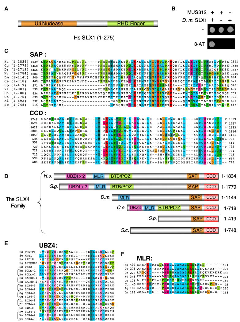
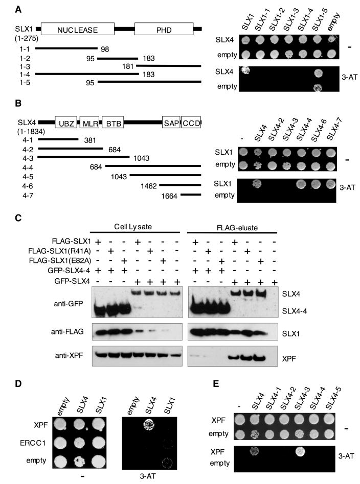
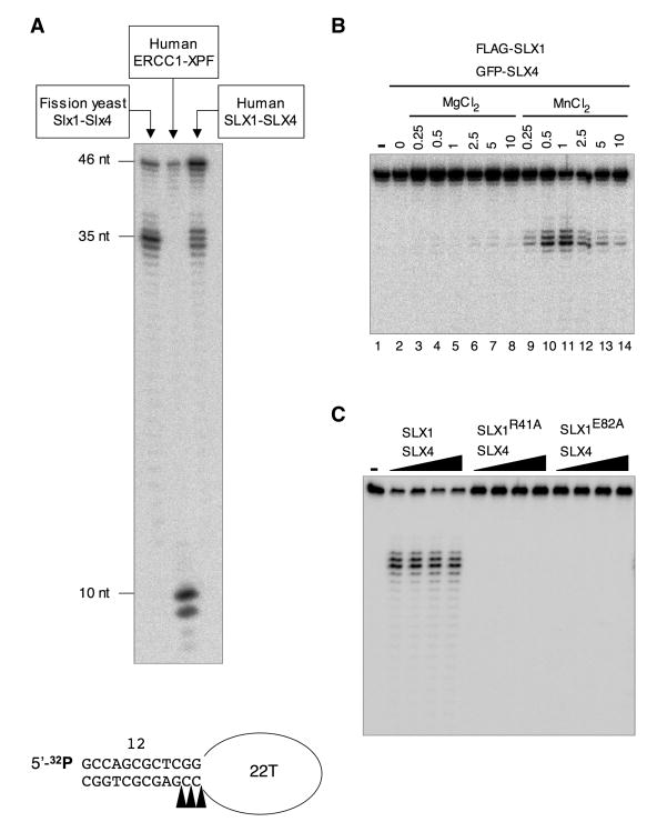
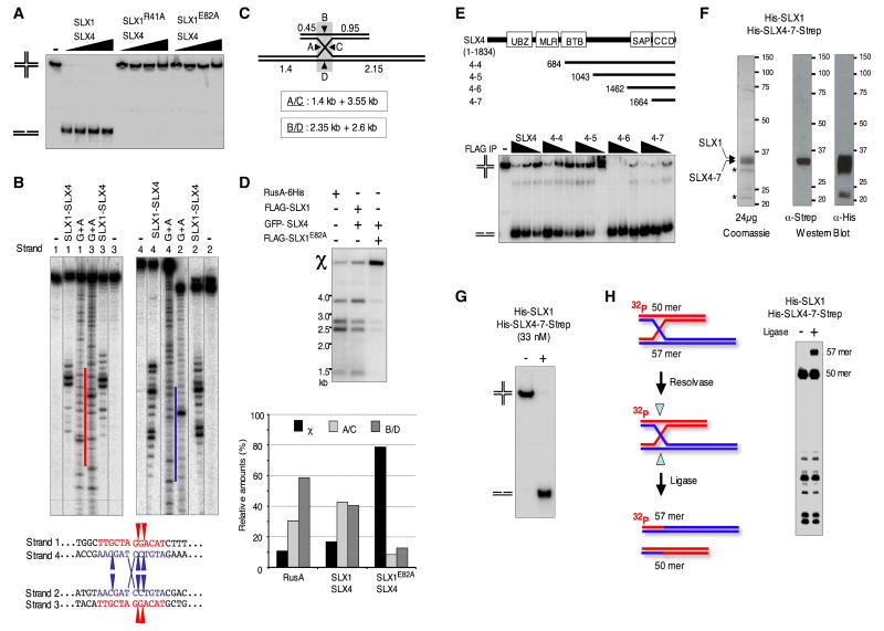
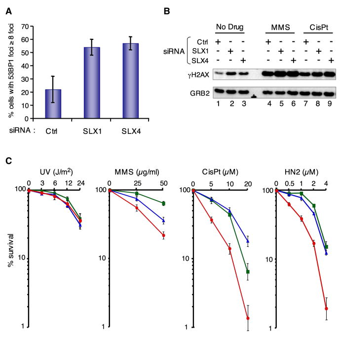
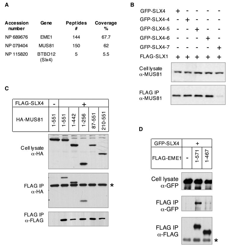
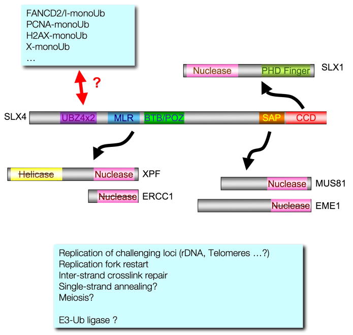
Structure-specific endonucleases resolve DNA secondary structures generated during DNA repair and recombination. The yeast 5' flap endonuclease Slx1-Slx4 has received particular attention with the finding that Slx4 has Slx1-independent key functions in genome maintenance. Although Slx1 is a highly conserved protein in eukaryotes, no orthologs of Slx4 were reported other than in fungi. Here we report the identification of Slx4 orthologs in metazoa, including fly MUS312, essential for meiotic recombination, and human BTBD12, an ATM/ATR checkpoint kinase substrate. Human SLX1-SLX4 displays robust Holliday junction resolvase activity in addition to 5' flap endonuclease activity. Depletion of SLX1 and SLX4 results in 53BP1 foci accumulation and H2AX phosphorylation as well as cellular hypersensitivity to MMS. Furthermore, we show that SLX4 binds the XPF(ERCC4) and MUS81 subunits of the XPF-ERCC1 and MUS81-EME1 endonucleases and is required for DNA interstrand crosslink repair. We propose that SLX4 acts as a docking platform for multiple structure-specific endonucleases.


Figures

FIGURE 1
FIGURE 1. The SLX4 family of proteins
A- schematic representation of SLX1 B- Y2H analysis of interactions between MUS312 fused to the GAL4 activation domain (AD) and D. melanogaster SLX1 fused to the GAL4 DNA binding domain (DBD). Strains were plated on selective media without or with 100 mM 3-AT. C- Sequence alignment of the SAP and CCD region of the following SLX4 metazoan proteins: Homo sapiens (H.s.), Gallus gallus (G.g.), Danio rerio (D.r.), Nematostella vectensis (N.v.), Drosophila melanogaster (D.m.), Caenorhabditis elegans (C.s.) and the following yeast: Schizosaccharomyces pombe (S.p.), Yarrowia lipolytica (Y.i.), Debaryomyces hansenii (D.h.), Candida albicans (C.a.), Kluyveromyces lactis (K.l.) and Saccharomyces cerevisiae (S.c.). Residues were colored using similarity groupings as follows: RED polar hydrophilic positively charged (K, R), DARK CYAN aromatic polar hydrophobic (H, Y), GREEN polar hydrophobic neutral (S, T, Q, N), BLUE hydrophobic non polar (A, I, L, M, P, W, V), FUSCHIA polar hydrophilic negatively charged (E, D), OLIVE (P), ORANGE tiny aliphatic neutral (G), PINK conserved C residues in UBZ4 motifs. D- Schematic representation of members of the SLX4 family. E- Sequence alignment of the UBZ4 motifs of the indicated SLX4 and DNA damage response proteins. F- Sequence alignment of the conserved motif located within the MUS312-MEI9 interaction-Like Region (MLR). Color coding is as in C-except for the conserved C residues in the UBZ4 highlighted in PINK. Tribolium castaneum (Tc.) and Anopheles gambiae (A.g.) were included in the alignment. Note: All accession numbers are listed in Supplemental data.
FIGURE 2
FIGURE 2. Human SLX4/MUS312 interacts with SLX1 and XPF
A- Y2H analysis of interactions between full length SLX4 fused to the GAL4-AD and full length or truncated SLX1 fused to the GAL4-DBD. B- Y2H analysis of interactions between full length SLX1 fused to GAL4-DBD and various SLX4 truncations fused to GAL4-AD. C- Immunoprecipitation of the indicated FLAG-tagged SLX1 proteins overproduced in HEK cells along with the indicated GFP-tagged SLX4 proteins. Western blot shows FLAG-tagged SLX1, GFP-tagged SLX4 and endogenous XPF in total cell lysates and FLAG-eluates. The amount of total cell lysate loaded per lane is 10% of the input, relative to the amount loaded of the corresponding FLAG-eluate. D- Y2H analysis of interactions between SLX4 and SLX1 fused to GAL4-AD and XPF and ERCC1 fused to GAL4-DBD. E- Y2H analysis of interaction between XPF fused to GAL4-DBD and the indicated SLX4 fragments fused to GAL4-AD.
FIGURE 3
FIGURE 3. Characterization of the 5′-flap endonuclease activity of human SLX1-SLX4
A- A 5′ 32P-end labeled stem loop substrate (SL) was incubated for 30 minutes at 30°C with 5 μl S. pombe TEV-Slx1 (Coulon et al., 2004), 150 ng rXPF-ERCC1 or 60 nl FLAG-eluate derived from HEK cells over-producing FLAG-SLX1 and GFP-SLX4. Reaction products were analyzed by denaturing PAGE. The cleavage sites are indicated on the schematized SL. B- SL was incubated with 60 nl FLAG-eluate (FLAG-SLX1/GFP-SLX4) for 5 min at 30°C in the presence of Mg++ or Mn++. C- The activity of 30, 60, 120, 240 nl FLAG-eluate derived from HEK cells over-producing FLAG-tagged wild type (WT) or the indicated mutant SLX1 proteins along with GFP-tagged SLX4 was compared on the SL substrate.
FIGURE 4
FIGURE 4. Human SLX1-SLX4 displays a remarkable HJ processing activity
A- An X12 HJ was incubated with FLAG-eluate derived from HEK cells over-producing FLAG-tagged WT or the indicated mutant SLX1 proteins along with GFP-tagged SLX4. Reactions were carried out side by side with those shown in Figure 3C under the exact same conditions. B- Four different X12 structures, each 5′ 32P-end labeled on a different strand, were incubated with FLAG-eluate (FLAG-SLX1/GFP-SLX4). Cleavage products from each X12 structure were analyzed by denaturing PAGE and run alongside an A+G Maxam-Gilbert sequence ladder derived from the strand labeled in the X12 structure. The homologous core is highlighted by red and blue color bars on the sequencing ladders. The position of the major incisions made in the homologous core (in red for strands 1 and 3 and in blue for strands 2 and 4), are indicated on the schematic. Note: Reactions with a strand 4-labeled X12 were performed on an asymmetric X12 junction (depicted in H-) where strands 1 and 4 are 57 nt long instead of 50. C- Schematic representation of the χ structure and the size of the expected resolution products following the A/C or B/D orientations. D- The χ structure was incubated with recombinant C-terminally 6His-tagged RusA (10nM) or 60 nl of the indicated FLAG-eluate for 40 min at 37°C. Relative amounts of the cleavage products generated after resolution of the χ structure following the A/C or B/D orientations and the remaining proportion of unresolved χ structure are plotted below the gel. E- Comparison of the activity on X12 of the indicated FLAG-eluate containing FLAG-tagged SLX1 and GFP-tagged full or C-terminal SLX4 fragments. Note: Western blot analysis of FLAG-tagged and GFP-tagged protein levels in the different FLAG-eluates is shown in Figure S10. F- Coomassie-blue stained proteins and Western blots showing the SLX1-SLX4-7 recombinant complex purified from E. coli. G- X12 was incubated 15 min at 30°C with recombinant human SLX1-SLX4-7 (33nM) produced in E. coli. H- Ligation assay of resolution products generated by recombinant SLX1-SLX4-7 shown in G-. An X12-L asymmetric junction containing 57 nt long strands 1 and 4 and 50 nt long strands 2 and 3, 5′ 32P-end labeled on strand 2, was incubated with SLX1-SLX4-7 (10nM) for 2.5 min at 30°C in the presence of 40 units of T4 DNA Ligase.
FIGURE 5
FIGURE 5. SLX1 and SLX4 are involved in preventing spontaneous and drug induced genome instability
A- MRC5 cells transfected with the indicated siRNA were stained with α-53BP1. 53BP1 foci formation was analyzed by confocal microscopy. Experiments were done in triplicate and the mean and standard deviation is shown. B- H2AX phosphorylation in mock-depleted, SLX1- or SLX4-depleted HEK 293 cells was analyzed by western blot using an α-γH2AX antibody. GRB2 detected with an α-GRB2 was used as a loading control. C- Hela cells were transfected with the indicated scrambled (green ■), SLX1 (blue ▲) or SLX4 (red ●) mRNA targeting siRNAs. MMS, cisPt, HN2 and UV-C treatments were done in triplicate and the mean and standard deviation is shown.
FIGURE 6
FIGURE 6. SLX4 associates with MUS81-EME1
A- Table summarizing the number of peptides and the percentage of coverage obtained by Mass Spectrometric analysis of a FLAG-EME1 precipitate. B- Western blot detection of endogenous MUS81 in total cell lysates of HEK cells co-producing FLAG-SLX1 and GFP-tagged full length or C-terminal SLX4 fragments and the corresponding FLAG-eluates. These are the same total cell lysates and FLAG-eluates than the ones described in Figure S10. The FLAG-eluates were also used in the nuclease assay shown in Figure 4E. The amount of total cell lysate loaded per lane is 10% of the input relative to the amount loaded of the corresponding FLAG-eluate. C- and D- HEK cells were transfected with pcDNA3 constructs expressing the indicated deletion constructs of MUS81 or EME1 and full length SLX4. Cell lysate and α-FLAG immunoprecipitates (IP) were analyzed by western blot with the indicated antibodies.
FIGURE 7
FIGURE 7
Speculative model describing SLX4 as a docking platform required for the coordination of structure-specific endonucleases in various genome maintenance pathways. The barred domains indicate functional domains that have diverged and lost key residues for the corresponding activity.

Comment in

References

    1. Aravind L, Koonin EV. SAP - a putative DNA-binding motif involved in chromosomal organization. Trends Biochem Sci. 2000;25:112–114. - PubMed
    1. Aravind L, Walker DR, Koonin EV. Conserved domains in DNA repair proteins and evolution of repair systems. Nucleic Acids Res. 1999;27:1223–1242. - PMC - PubMed
    1. Boddy MN, Gaillard PH, McDonald WH, Shanahan P, Yates JR, Russell P. Mus81-Eme1 are essential components of a Holliday junction resolvase. Cell. 2001;107:537–548. - PubMed
    1. Bolt EL, Lloyd RG. Substrate specificity of RusA resolvase reveals the DNA structures targeted by RuvAB and RecG in vivo. Mol Cell. 2002;10:187–198. - PubMed
    1. Brookman KW, Lamerdin JE, Thelen MP, Hwang M, Reardon JT, Sancar A, Zhou ZQ, Walter CA, Parris CN, Thompson LH. ERCC4 (XPF) encodes a human nucleotide excision repair protein with eukaryotic recombination homologs. Mol Cell Biol. 1996;16:6553–6562. - PMC - PubMed

Publication types