. 2009 Jul 10;138(1):146-59.
doi: 10.1016/j.cell.2009.04.041.

CarD is an essential regulator of rRNA transcription required for Mycobacterium tuberculosis persistence

Affiliations

CarD is an essential regulator of rRNA transcription required for Mycobacterium tuberculosis persistence

Christina L Stallings et al. Cell. .

Abstract

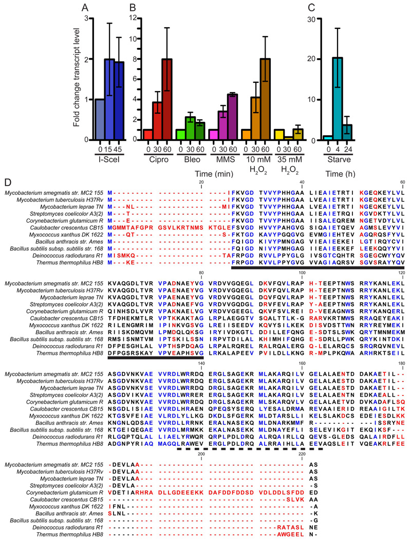
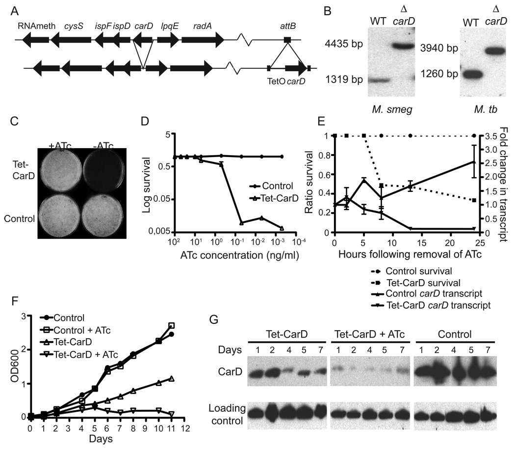
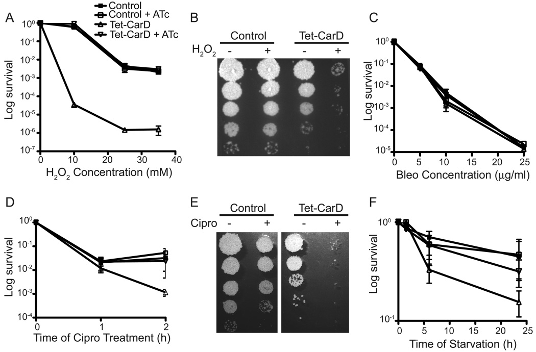
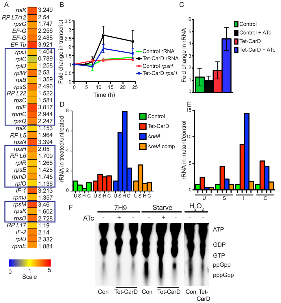
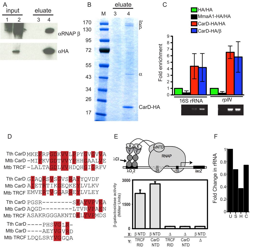
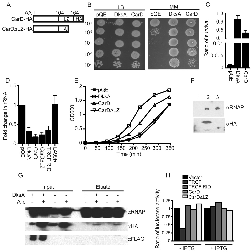
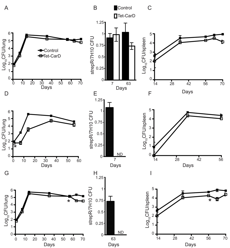
Mycobacterium tuberculosis is arguably the world's most successful infectious agent because of its ability to control its own cell growth within the host. Bacterial growth rate is closely coupled to rRNA transcription, which in E. coli is regulated through DksA and (p)ppGpp. The mechanisms of rRNA transcriptional control in mycobacteria, which lack DksA, are undefined. Here we identify CarD as an essential mycobacterial protein that controls rRNA transcription. Loss of CarD is lethal for mycobacteria in culture and during infection of mice. CarD depletion leads to sensitivity to killing by oxidative stress, starvation, and DNA damage, accompanied by failure to reduce rRNA transcription. CarD can functionally replace DksA for stringent control of rRNA transcription, even though CarD associates with a different site on RNA polymerase. These findings highlight a distinct molecular mechanism for regulating rRNA transcription in mycobacteria that is critical for M. tuberculosis pathogenesis.


Figures

Figure 1
Figure 1. carD transcription is upregulated in response to oxidative stress, DNA damage, and starvation
carD transcript levels in treated log phase wild-type M. smegmatis cultures were measured by qRT-PCR, normalized to sigA transcript levels, and expressed as a fold change from untreated cultures. Each experiment was done in triplicate. Graphical data in this and subsequent figures are represented as mean ± SEM. (A) carD transcript levels during DSBs. I-SceI expression is induced at time 0. (B) carD transcript levels during genotoxic stress. M. smegmatis Mc2155 was treated with 10 µg/ml Ciprofloxacin (Cipro), 10 µg/ml bleomycin (bleo), 0.1% MMS, and 10 or 35 mM H2O2. (C) carD transcript levels during nutrient deprivation. M. smegmatis was washed once and starved in PBS+0.05% Tween 80. (D) Amino acid sequence alignment of selected CarD proteins. Letter coloring indicates the sequence conservation. Blue is highly conserved (>66%) and red is not conserved (<33%). The solid black line designates the region of CarD that is homologous to the TRCF RID. The dashed black line designates the leucine zipper motif.
Figure 2
Figure 2. CarD is essential for growth of M. tuberculosis and M. smegmatis in culture
(A) Diagram of the carD gene region in M. smegmatis and M. tuberculosis and the construction of M. smegmatis ΔcarD attb∷tetcarD. Arrangement of M. tuberculosis ΔcarD attb∷tetcarD is identical except a hygR cassette replaces carD. (B) Southern blot analysis of wild-type and ΔcarD attb∷tetcarD M. smegmatis (left panel) and M. tuberculosis (right panel). Wild-type M. smegmatis yields a 1319 bp band, while ΔcarD results in a 4435 bp band. Wild-type M. tuberculosis yields a 1260 bp band, while ΔcarD results in a 3940 bp band. (C and D) Growth of M. smegmatis Tet-CarD (mgm1703) and control (mgm1701) strains on LB plates containing varying concentrations of ATc. (C) shows the growth of Tet-CarD and the control strain on plates containing either 50 ng/ml ATc or no ATc. (D) shows the ratio of survival of each strain on different concentrations of ATc as compared to in the presence of 50 ng/ml. (E) Survival and carD transcript levels of M. smegmatis Tet-CarD and the control strain following removal of ATc. Each strain was diluted to an OD600 of 0.05 into media either with or without ATc. At each time point survival was determined by plating dilutions onto LB + 50 ng/ml ATc and is expressed as a ratio to CFUs from the respective culture in 50 ng/ml ATc (left axis, dashed lines). qRT-PCR was performed on RNA samples collected at each time point and carD transcript levels were normalized to sigA transcript levels and expressed as a fold change from time 0 (right axis, solid lines). (F) Growth of M. tuberculosis Tet-CarD (mgm1797) and control strain (mgm1799) in 7H9 broth with or without ATc. Each strain was diluted to an OD600 of 0.05 into the appropriate media and the OD600 was measured at the designated time points. In this system, ATc represses carD expression. (G) Western blot analysis of total protein lysates of the same cultures in (F) using rabbit polyclonal antibodies specific for CarD and DlaT (loading control).
Figure 3
Figure 3. CarD is required for resistance to H2O2, Ciprofloxacin, and nutrient deprivation
(A–E) Survival of M. smegmatis strains during genotoxic stress. The strain symbols listed in the legend in (A) are the same for all panels. Tet-CarD (mgm1703) and the control strain (mgm1701) growing in LB broth with or without ATc were treated for 1 h with varying concentrations of H2O2 (A–B), bleomycin (C), or treated for 1 and 2 hours with 10µg/ml of Ciprofloxacin (D–E). Following treatment, dilutions were plated on LB + ATc and survival is expressed as a ratio of CFUs compared to untreated cultures. (B and E) display the plating efficiency of Tet-CarD and the control strain grown in the absence of ATc after treatment with 10 mM H2O2 for 1 h (B) and 10µg/ml Ciprofloxacin for 2 h (E). (F) Survival of M. smegmatis strains during nutrient deprivation. Tet-CarD and control strains in LB with or without ATc were washed and starved in PBS + 0.05% Tween 80 for the indicated times. Survival was determined and expressed as described above.
Figure 4
Figure 4. CarD is required for the stringent response
(A) Heat map of upregulated ribosomal proteins and translation factors (p < 0.05) during CarD depletion in M. smegmatis. Microarray analyses compared M. smegmatis Tet-CarD (mgm1703) to the control strain (mgm1701) 13 hours after CarD depletion in LB media. The scale is shown at the bottom, gene names are listed to the left and fold changes in expression during CarD depletion compared to the control strain are to the right. Predicted operons are boxed. (B) 16S rRNA and rpsH transcript levels in M. smegmatis strains from Figure 2E during growth in the absence of ATc, as determined by qRT-PCR and expressed as a fold change from time 0. (C) 16S rRNA levels in M. tuberculosis strains from Figure 2F in the presence and absence of ATc, as determined by qRT-PCR. The fold change in rRNA levels at 7 days compared to day 0 is graphed. (D and E) 16S rRNA levels in M. smegmatis control, Tet-CarD, ΔrelA, and ΔrelA complemented with a WT relA gene (ΔrelA comp) strains during starvation, oxidative, and genotoxic stress. Each strain was diluted to an OD600 of 0.05 into LB - ATc. Cultures were then left untreated (U), starved in PBS + 0.05% Tween 80 for 4 h (S), treated for 1 h with 10 mM H2O2 (H), or 2 h with 10µg/ml Ciprofloxacin (C) before collecting samples for qRT-PCR analysis of 16S rRNA. (D) shows the fold change of rRNA in each strain during treatment as compared to untreated (set to 1). Using the same data as in (D), (E) illustrates the rRNA levels in each mutant compared to the CarD+ control strain in the same conditions. In all assays, rpsH and rRNA transcript levels were normalized to sigA transcript levels. (F) (p)ppGpp accumulation during the stringent response. M. smegmatis Tet-CarD and control strain (Con) were diluted to an OD600 of 0.05 in 7H9 broth with or without ATc for 10 h before labeling intracellular nucleotides in untreated cultures (7H9) or during nutrient deprivation (starve) and oxidative stress (H2O2) and analyzing by TLC. The migration positions of ATP, GTP, and ppGpp were determined by nonradioactive nucleotide controls.
Figure 5
Figure 5. CarD interacts directly with the N-terminus of the RNAP β subunit
(A–B) Immunoprecipitation experiments with HA antibody in either M. smegmatis ΔcarD attb∷tetcarD (lanes 1 and 3) or M. smegmatis ΔcarD attB∷tetcarD-HA (lanes 2 and 4). (A) Inputs and eluates analyzed by western blot analysis using antibodies specific for either the RNAP β subunit or HA. (B) Eluates analyzed by coomassie blue staining. Co-precipitated RNAP β, β’, and α subunits were identified by MALDI-TOF-MS. (C) CarD-HA and RNAP β coprecipitated fragments of DNA containing the promoters of the rRNA and rplN operons. ChIP assays were performed using antibodies specific for HA on exponentially growing M. smegmatis ΔcarD attb∷tetcarD-HA (CarD-HA/HA), M. smegmatis expressing HA alone (HA/HA), or M. smegmatis expressing MmaA1-HA (MmaA1-HA/HA) as well as using antibodies specific for RNAP β with ΔcarD attB∷tetcarD-HA cultures (CarD-HA/β). Precipitated and input DNA corresponding to the promoter regions of the rRNA and the rplN operon was determined by quantitative PCR. The graph shows the fold enrichment of DNA product compared to the amount of product precipitated from the strain expressing HA alone and agarose gel electrophoresis of PCR products. MmaA1 is a mycolic acid methyltransferase that does not interact with DNA and served as a negative control. (D) Amino acid sequence alignment of T. thermophilus (Tth) CarD amino acids 1–60, M. tuberculosis (Mtb) CarD residues 1–60, and the M. tuberculosis TRCF RID (amino acids 516–587). (E) T. thermophilus CarD and TRCF interact with the N-terminus of the RNAP β subunit. Cartoon depicts how contact between a protein domain (X) fused to the α subunit of E. coli RNAP and a partner domain (Y) fused to a DNA-binding protein (the CI protein of bacteriophage λ) activates transcription from test promoter placOL2–62, which bears an upstream recognition site for λCI (λOL2). In reporter strain FW102 OL2–62 (Deaconescu et al., 2006), test promoter placOL2–62 is located on an F' episome and drives the expression of a linked lacZ gene. For these experiments, the N-terminus of β (residues 10–133) was fused to α and the TRCF RID (residues 314–444) or N-terminus of CarD (residues 1–66) was fused to λCI. The bar graph shows the results of β-galactosidase assays (mean and SEM of four independent determinations) performed with FW102 OL2–62 cells that contained two compatible plasmids; one that encoded the α–β fusion protein or wild-type α (Δ), and another that encoded the λCI-TRCF RID fusion protein, the λCI-CarD NTD fusion protein or wild-type λCI (Δ). (F) M. smegmatis Δmfd is not deficient in stringent control. M. smegmatis Δmfd was left untreated (U), starved in PBS + 0.05% Tween 80 for 4 h (S), treated for 1 h with 10 mM H2O2 (H), or 2 h with 10µg/ml Ciprofloxacin (C) before collecting samples for RNA extraction and qRT-PCR analysis of 16S rRNA. The fold change of rRNA is compared to untreated (set to 1).
Figure 6
Figure 6. CarD can functionally replace DksA but not TRCF
(A) Illustration of CarD-HA and the truncation deleting the CarD leucine zipper (CarDΔLZ-HA) (B and C) Survival of ΔdksA complemented strains on minimal media. Dilutions of ΔdksA E. coli strains expressing DksA, CarD-HA (CarD), or vector (pQE) were plated on either LB or M9 minimal media (MM). Survival was determined by the ratio of CFUs on minimal media compared to LB. (D) rRNA levels in ΔdksA complemented strains. E. coli ΔdksA and the indicated complemented strains were diluted into M9 minimal media for 2 h. 16S rRNA levels in each strain as determined by qRT-PCR, normalized to transcripts from the β-lactamase gene (bla) in pQE-30, and expressed as a fold change compared to levels in ΔdksA harboring the vector alone. (E) Growth curve of ΔdksA complemented strains in LB. (F) CarD associates with the E. coli RNAP. Immunoprecipitation experiments with HA antibody matrix of cell extracts prepared from log phase E. coli ΔdksA strains containing the vector alone (lane 1) or expressing either CarD-HA (lane 2) or CarDΔLZ-HA (lane 3). Eluates were analyzed by immunoblotting using anti-RNAP β or HA antibodies. (G) DksA does not interact with the mycobacteria RNAP. M. smegmatis ΔcarD attB∷tetcarD-HA and M. smegmatis ΔcarD attB∷tetcarD-HA expressing FLAG-DksA were diluted to an OD600 of 0.05 in LB broth with or without ATc for 13 h before performing immunoprecipitation experiments with antibodies specific for the RNAP β subunit. Inputs and eluates were analyzed by western blot analyses using antibodies specific for RNAP β, HA, and FLAG. (H) CarD cannot replace TRCF in the roadblock repression assay. E. coli UNCNOMFD (Δmfd) with the roadblock repression luciferase reporter construct (pRCB-KA4) and an empty pET21a vector or pET21a plasmids encoding the indicated proteins. Luciferase activities in the presence and absence of 0.5 mM IPTG shown are the average of three independent experiments and are expressed as a fraction of luciferase activity in cells harboring the empty pET21a vector (set to 1).
Figure 7
Figure 7. CarD is necessary for replication and persistence of M. tuberculosis in mice
Bacterial titers in the lungs (A, D, G) and spleens (C, F, I) of C57Bl/6 mice infected with either mgm1799 (control) or mgm1797 (Tet-CarD), both carrying the revTetR on a strepR plasmid. The strain symbols listed in the legend for (A) and (B) are the same for all panels. (A–C) were given regular drinking water throughout the infection, (D–F) were administered doxycycline in their drinking water starting on day 1 (designated by the star), and (G–I) were given doxycycline in their drinking water starting on day 56 (designated by the star). (B, E, H) show the ratio of CFUs from the lung grown on 7H10 plates containing streptomycin as compared to 7H10 containing no antibiotics. ND denotes when no colonies were detected after plating 4% of the lung homogenate. Data are means ± SEM of four mice per group and time point from one of two replicate experiments.

References

    1. Alfoldi L, Stent GS, Clowes RC. The chromosomal site of the RNA control (RC) locus in Escherichia coli. J Mol Biol. 1962;5:348–355. - PubMed
    1. Avarbock D, Avarbock A, Rubin H. Differential regulation of opposing RelMtb activities by the aminoacylation state of a tRNA.ribosome.mRNA.RelMtb complex. Biochemistry. 2000;39:11640–11648. - PubMed
    1. Avarbock D, Salem J, Li LS, Wang ZM, Rubin H. Cloning and characterization of a bifunctional RelA/SpoT homologue from Mycobacterium tuberculosis. Gene. 1999;233:261–269. - PubMed
    1. Betts JC, Lukey PT, Robb LC, McAdam RA, Duncan K. Evaluation of a nutrient starvation model of Mycobacterium tuberculosis persistence by gene and protein expression profiling. Mol Microbiol. 2002;43:717–731. - PubMed
    1. Boshoff HI, Myers TG, Copp BR, McNeil MR, Wilson MA, Barry CE., 3rd The transcriptional responses of Mycobacterium tuberculosis to inhibitors of metabolism: novel insights into drug mechanisms of action. J Biol Chem. 2004;279:40174–40184. - PubMed

Publication types

MeSH terms

Substances

Associated data