

Hepatocyte GP73 expression in Wilson disease
- PMID: 19596473
- PMCID: PMC2750828
- DOI: 10.1016/j.jhep.2009.05.029
Hepatocyte GP73 expression in Wilson disease
Abstract
Background/aims: Wilson disease (WD) is a disorder of copper transport caused by mutations within the ATP7B gene. WD is phenotypically variable and can present with predominantly hepatic or neurologic manifestations. The mechanisms responsible for this variability are unknown. GP73, a Golgi membrane protein, is expressed in hepatocytes in response to acute and chronic liver disease.
Methods: Hepatocyte GP73 expression was examined in the livers of WD patients by semiquantitative immunohistochemistry. GP73 mRNA levels were measured in mice with a deletion of the WD gene (Atp7b(-/-)) by real-time PCR, and these values were compared to the concomitant histological abnormalities and previously reported copper levels.
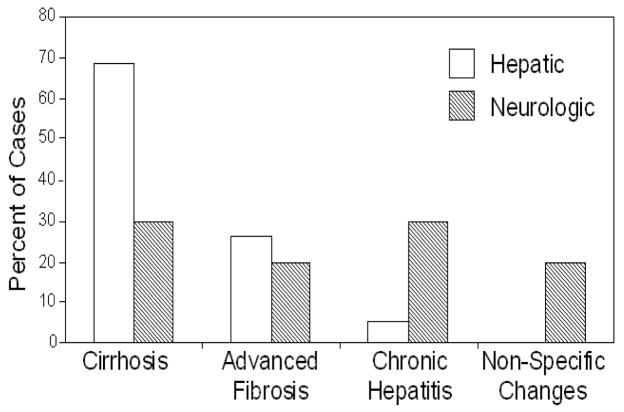
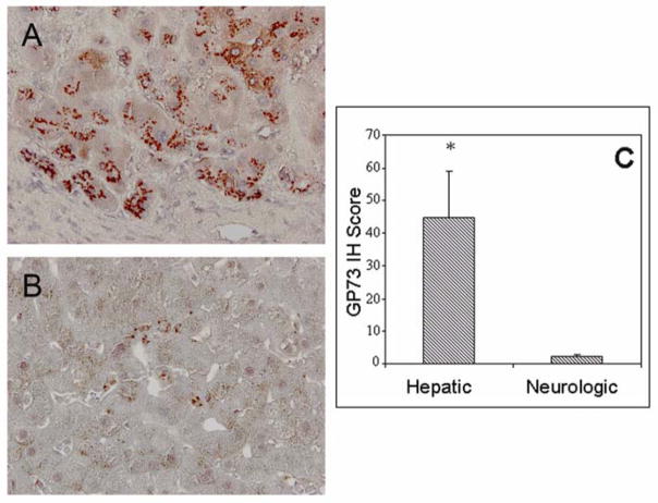
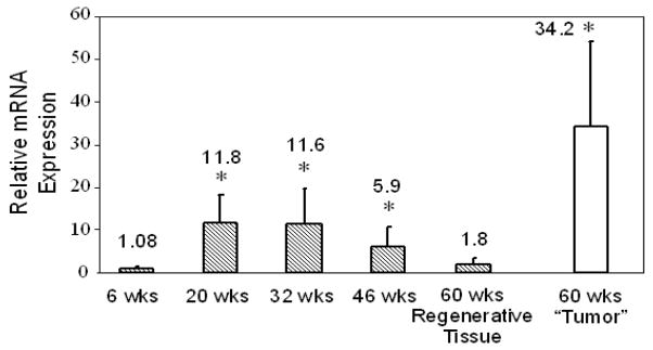
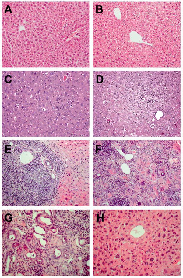
Results: Hepatocyte GP73 expression was more frequently observed in patients with hepatic versus neurologic presentation (79% vs. 30%, p<0.05). Furthermore, GP73 expression was significantly higher (44.7+/-14.0 vs. 2.0+/-0.81, p<0.05) in patients with hepatic phenotype. In Atp7b(-/-) mice, GP73 mRNA was significantly elevated at 20-46 weeks of age, coincident with extensive hepatic inflammation and fibrosis, but not at 6 weeks, when hepatic histology was normal despite significant copper overload. GP73 mRNA levels normalized concomitantly with the resolution of hepatic injury at 60-weeks. However, in tumor-like nodules GP73 was strikingly elevated.
Conclusion: Increased hepatocyte GP73 expression is more commonly a feature of hepatic than neurologic WD, and is triggered in response to inflammation, fibrosis, and dysplasia, rather than copper overload.
Figures
References
-
- Iftikhar R, Kladney RD, Havlioglu N, Schmitt-Graff A, Gusmirovic I, Solomon H, et al. Disease- and cell-specific expression of GP73 in human liver disease. Am J Gastroenterol. 2004;99:1087. - PubMed
-
- Kladney RD, Cui X, Bulla GA, Brunt EM, Fimmel CJ. Expression of GP73, a resident Golgi membrane protein, in viral and nonviral liver disease. Hepatology. 2002;35:1431. - PubMed
-
- Bachert C, Fimmel C, Linstedt AD. Endosomal trafficking and proprotein convertase cleavage of cis Golgi protein GP73 produces marker for hepatocellular carcinoma. Traffic. 2007;8:1415. - PubMed
-
- Marrero JA, Romano PR, Nikolaeva O, Steel L, Mehta A, Fimmel CJ, et al. GP73, a resident Golgi glycoprotein, is a novel serum marker for hepatocellular carcinoma. J Hepatol. 2005;43:1007. - PubMed
Publication types
MeSH terms
Substances
Grants and funding
LinkOut - more resources
Full Text Sources
Medical
Molecular Biology Databases
Miscellaneous
