. 2009 Sep;29(18):4918-34.
doi: 10.1128/MCB.00009-09. Epub 2009 Jul 13.

Arginine methylation regulates telomere length and stability

Affiliations

Arginine methylation regulates telomere length and stability

Taylor R H Mitchell et al. Mol Cell Biol. 2009 Sep.

Abstract

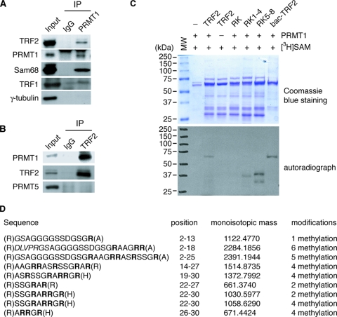
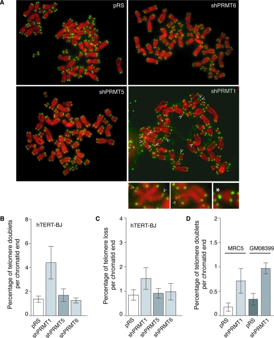
TRF2, a component of the shelterin complex, functions to protect telomeres. TRF2 contains an N-terminal basic domain rich in glycines and arginines, similar to the GAR motif that is methylated by protein arginine methyltransferases. However, whether arginine methylation regulates TRF2 function has not been determined. Here we report that amino acid substitutions of arginines with lysines in the basic domain of TRF2 induce telomere dysfunction-induced focus formation, leading to induction of cellular senescence. We have demonstrated that cells overexpressing TRF2 lysine mutants accumulate telomere doublets, indicative of telomere instability. We uncovered that TRF2 interacts with PRMT1, and its arginines in the basic domain undergo PRMT1-mediated methylation both in vitro and in vivo. We have shown that loss of PRMT1 induces growth arrest in normal human cells but has no effect on cell proliferation in cancer cells, suggesting that PRMT1 may control cell proliferation in a cell type-specific manner. We found that depletion of PRMT1 in normal human cells results in accumulation of telomere doublets, indistinguishable from overexpression of TRF2 lysine mutants. PRMT1 knockdown in cancer cells upregulates TRF2 association with telomeres, promoting telomere shortening. Taken together, these results suggest that PRMT1 may control telomere length and stability in part through TRF2 methylation.


Figures

FIG. 1.
FIG. 1.
Arginines in the basic domain of TRF2 are crucial for its function. (A) Schematic diagram of human TRF2. Arginines in the basic domain are highlighted in red, and they are conserved between human and mouse. (B) Western blot analysis of expression of the wild type and various TRF2 mutants. Whole-cell extracts made from 200,000 cells were used, and immunoblotting was performed with anti-TRF2 antibody. The γ-tubulin blot was used as a loading control. (C) Growth curve of HT1080 cells expressing TRF2 carrying amino acid changes of arginines to lysines or alanines. Cells were infected with retrovirus expressing either wild-type TRF2, various TRF2 mutants, or the vector alone. Following the last infection, cells were selected with puromycin (2 μg/ml) and maintained in the selection medium for 13 days. (D) Growth curve of hTERT-BJ cells expressing TRF2 carrying amino acid changes of arginines to lysines. Cells were infected with the indicated retrovirus. Following the last infection, cells were selected with puromycin (2 μg/ml) and maintained in the selection medium for 15 days. Standard deviations derived from three independent experiments are indicated. (E) Overexpression of TRF2 carrying amino acid changes of arginines to lysines induces senescence. hTERT-BJ cells infected with the indicated viruses were stained for senescence-associated β-Gal on day 13. (F) Quantification of percentage of cells staining positive for senescence-associated β-Gal. A total of more than 1,500 cells from three independent experiments were scored. Standard deviations derived from three independent experiments are indicated.
FIG. 2.
FIG. 2.
(A) Overexpresssion of TRF2-RK induces TIF formation. Indirect immunofluorescence using anti-TRF1 in conjunction with anti-53BP1 was performed with fixed hTERT-BJ cells expressing either TRF2-RK or the vector alone. Arrowheads indicate sites of colocalization of 53BP1 and TRF1. (B) Quantification of percentage of cells with more than five TIFs. For each cell line, a total of more than 800 cells from three independent experiments were scored. Standard deviations derived from three independent experiments are indicated. (C) Dot blots of ChIPs. ChIPs were performed with either anti-TIN2 or anti-IgG antibody in cell extracts from HT1080 cells overexpressing TRF2-RK or the vector alone. Precipitated DNA was analyzed for the presence of TTAGGG repeats and Alu repeats by Southern blotting. (D) Quantification of anti-TIN2 ChIPs. The signals were quantified by ImageQuant analysis. The percentage of precipitated DNA was calculated relative to the input signals. Standard deviations derived from three independent experiments are indicated. (E) Western blot analysis of TRF2 protein expression. Whole-cell extracts made from 200,000 cells were used, and immunoblotting was performed with anti-TRF2 and anti-TIN2 antibodies. The γ-tubulin blot was used as a loading control. (F) Genomic blot of telomeric restriction fragments of hTERT-BJ cells expressing various proteins as indicated above the blot. About 3 μg of RsaI/HinfI-digested genomic DNA from each sample was used for gel electrophoresis. The DNA molecular size markers are shown on the left of the blot. The bottom panel, taken from an ethidium bromide-stained agarose gel, is used as a loading control. (G) Genomic blot of telomeric restriction fragments of hTERT-BJ cells expressing various proteins as indicated above the blot. About 3 μg of RsaI/HinfI-digested genomic DNA from each sample was separated on a CHEF gel. The picture of an ethidium bromide (EtBr)-staining gel is shown at the right of the blot, whereas the DNA molecular size markers are shown at the left of the blot.
FIG. 3.
FIG. 3.
Overexpression of TRF2 carrying amino acid changes of arginines to lysines promotes the formation of telomere doublets in both hTERT-BJ and Nbs1-deficient GM07166 cells. (A) Metaphase spreads from hTERT-BJ cells expressing TRF2-RK, TRF2ΔB, or the vector alone. Chromosomes were stained with DAPI and false colored in red. Telomeric DNA was detected by FISH using a FITC-conjugated (CCCTAA)3-containing PNA probe (green). Arrows indicate telomere doublets. Enlarged images of chromosomes with telomere doublets are shown. (B) Quantification of telomere doublets in hTERT-BJ cells stably infected with retrovirus as indicated. For each cell line, a total of 1,600 to 1,900 chromosomes from 42 to 45 metaphase cells were scored. Standard deviations derived from at least three independent experiments are indicated. (C) Quantification of telomere loss in hTERT-BJ cells expressing TRF2 alleles as indicated. For each cell line, a total of 1,600 to 1,900 chromosomes from 42 to 45 metaphase cells were scored. Standard deviations derived from at least three independent experiments are indicated. (D) Quantification of telomere doublets in Nbs1-deficient GM07166 cells stably infected with retrovirus as indicated. For each cell line, a total of 1,372 to 1,650 chromosomes from 38 to 40 metaphase cells were scored. Standard deviations derived from three independent experiments are indicated.
FIG. 4.
FIG. 4.
Arginines of TRF2 at positions 17 and 18 are methylated in vivo. Liquid chromatography-tandem mass spectrometry analysis was performed with endogenous TRF2 immunoprecipitated from HeLa cells. The spectra of two peptides identified to contain methylated arginines in the basic domain are shown, with the relative abundance plotted against the monoisotopic mass (m/z). The m/z peaks from both b-type and y-type ions are indicated. The peptide presented in panel A contained dimethylated arginine at position 17 and monomethylated arginine at position 18, whereas the peptide presented in panel B contained dimethylated arginine at position 17.
FIG. 5.
FIG. 5.
Interaction of PRMT1 with TRF2. (A) Association of PRMT1 with TRF2 in vivo. Coimmunoprecipitations with HeLa nuclear extracts were conducted with either anti-IgG or anti-PRMT1 antibody. Western analysis was done with anti-PRMT1, anti-TRF2, anti-TRF1, anti-Sam68, and anti-γ-tubulin antibodies. (B) Association of TRF2 with PRMT1 in vivo. Coimmunoprecipitations with HeLa nuclear extracts were conducted with either anti-IgG or anti-TRF2 antibody. Western analysis was done with anti-PRMT1, anti-TRF2, and anti-PRMT5 antibodies. (C) PRMT1 methylates arginines in the basic domain of TRF2. In vitro methylation assays were conducted using [3H]SAM, GST-PRMT1, and recombinant TRF2 as indicated. Both the Coomassie blue-staining gel (top panel) and autoradiograph (bottom panel) are shown. The protein molecular mass markers are shown on the left of the gel and blot. All proteins except for bac-TRF2 were expressed in bacteria. Bac-TRF2 (a gift from Titia de Lange) refers to wild-type TRF2 derived from baculovirus. (D) PRMT1 methylates multiple arginines of the basic domain in vitro. Bacterium-expressed wild-type TRF2 was methylated in vitro and then subjected to matrix-assisted laser desorption ionization mass spectrometry analysis. Peptides containing methylated arginines are shown. Arginines in the basic domain are highlighted in bold.
FIG. 6.
FIG. 6.
PRMT1 is the main enzyme responsible for methylating TRF2 in vivo. (A) Affinity-purified 5510 antibody specifically recognizes TRF2 peptide containing asymmetrically dimethylated arginine 17 (R17). Increasing amounts of peptide carrying either unmodified R17 or asymmetrically dimethylated R17 were spotted onto a nitrocellulose membrane, followed by immunoblotting with affinity-purified 5510 or crude serum (5510CS). The amounts of peptide spotted, from left to right, are 0.5 μg, 1.3 μg, and 2.7 μg. (B) Peptide competition assays. Affinity-purified 5510 antibody was incubated with 5.5 μg of unmodified peptide prior to immunoblotting. Crude serum 5510 was used to show the presence of the TRF2 peptide on the nitrocellulose membrane. Amounts of peptide spotted, from left to right, for both modified and unmodified, are 0.5 μg, 1.3 μg, and 2.7 μg. (C) Peptide competition assays. Prior to immunoblotting TRF2 peptide carrying asymmetrically dimethylated R17, affinity-purified 5510 antibody was incubated with 5.5 μg of either unmodified or modified peptide as indicated. Crude serum 5510 was used to show the presence of the methylated TRF2 peptide. (D) Western analysis of methylated TRF2. Twenty micrograms of the whole-cell extract from hTERT-BJ cells was immunoblotted with affinity-purified 5510 antibody. (E) Depletion of endogenous TRF2 leads to loss of methylated TRF2 bound by 5510 antibody. hTERT-BJ cells were infected with retrovirus expressing either shTRF2 or the vector pRS alone. Western analysis was performed with 5510 and anti-TRF2. The γ-tubulin blot was used as a loading control. (F) Western analysis of methylated TRF2. Affinity-purified 5510 antibody was incubated with 5.5 μg of unmodified peptide prior to immunoblotting. Twenty micrograms of the whole-cell extract from several cell lines, as indicated, was used for immunoblotting. (G) 5510 recognizes immunoprecipitated TRF2. Endogenous TRF2 was immunoprecipitated from HeLa cells using anti-TRF2 antibody. Immunoblotting was performed with anti-TRF2 antibody or affinity-purified 5510 antibody that had been preincubated with 5.5 μg of unmodified peptide. (H) Depletion of endogenous PRMT1 decreases asymmetrical dimethylation of R17. hTERT-BJ, HT1080, and 293T cells were infected with retrovirus expressing either shPRMT1 or the vector pRS alone. For detection of methylated TRF2, Western analysis was done with 5510 that had been preincubated with 5.5 μg of unmodified peptide. Western analysis was also done with anti-TRF2 and anti-PRMT1. The γ-tubulin blot was used as a loading control. (I) Depletion of endogenous PRMT6 has no effect on dimethylation of R17. hTERT-BJ cells were infected with retrovirus expressing either shPRMT6 or the vector pRS alone. Western analysis was done with 5510, anti-TRF2, and anti-PRMT6. The γ-tubulin blot was used as a loading control.
FIG. 7.
FIG. 7.
Depletion of PRMT1 results in induction of growth arrest in normal human fibroblast cells. (A) Western analysis of PRMT1 expression. Whole-cell extracts made from 200,000 cells were used, and immunoblotting was performed with anti-PRMT1 antibody. The γ-tubulin blot was used as a loading control. (B) Western analysis of PRMT5 expression. Immunoblotting was conducted with anti-PRMT5 antibody, and the γ-tubulin blot was used as a loading control. (C) Western analysis of PRMT6 expression. Immunoblotting was conducted with anti-PRMT6 and anti-γ-tubulin antibodies, the latter serving as a loading control. (D) Growth curve of hTERT-BJ cells infected with indicated virus. hTERT-BJ cells expressing shPRMT1 undergo growth arrest within 3 days of infection, and seeding after infection was associated with massive cell loss. To overcome this problem, 4.8 × 10⁴ cells were seeded in triplicate on day −4. Cells were infected five times with indicated virus over 12-h intervals between day −3 and day −1. Selection with puromycin (2 μg/ml) started on day 0, and cells were maintained in the selection medium for 9 days. Standard deviations derived from three independent experiments are indicated. (E) Growth curve of normal primary fibroblast MRC5 cells infected with indicated virus. MRC5 cells expressing shPRMT1 undergo growth arrest. Seeding, retroviral infection, and selection of MRC5 were done as described for panel D. Standard deviations derived from three independent experiments are indicated. (F) Loss of PRMT1 results in growth arrest in normal human cells. Live cell images show hTERT-BJ, IMR90, or GM08399 cells infected with retrovirus expressing shPRMT1 or the vector pRS alone. Images were taken 5 days after infection. (G) Genomic blots of telomeric restriction fragments from hTERT-BJ, IMR90, and MRC5 cells expressing either shPRMT1 or pRS. About 3 μg of RsaI/HinfI-digested genomic DNA from each sample was used for gel electrophoresis. The DNA molecular size markers are shown to the left of the blots.
FIG. 8.
FIG. 8.
Depletion of PRMT1 in human normal cells results in telomere instability, promoting the formation of telomere doublets. (A) Analysis of metaphase chromosomes from hTERT-BJ cells infected with indicated virus. Chromosomes were stained with DAPI and false colored in red. Telomeric DNA was detected by FISH using a FITC-conjugated (CCCTAA)3-containing PNA probe (green). Arrows indicate telomere doublets, and the asterisk represents telomere loss. Enlarged images of chromosomes with telomere doublets are shown at the bottom. (B and C) Quantification of telomere doublets and telomere loss in hTERT-BJ cells stably infected with the indicated virus. For each cell line, a total of more than 2,700 chromosomes from at least 60 metaphase cells were scored. Standard deviations derived from at least three independent experiments are indicated. (D) Quantification of telomere doublets in MRC5 and GM08399 cells stably infected with the indicated virus. For each cell line, a total of more than 1,600 chromosomes from 40 to 44 metaphase cells were scored. Standard deviations derived from at least three independent experiments are indicated.
FIG. 9.
FIG. 9.
Depletion of PRMT1 does not affect cell proliferation in human cancer cells. (A) Western analysis of PRMT1 expression in HT1080, 293T, and GM637 cells expressing shPRMT1 or pRS. The γ-tubulin blot was used as a loading control. (B) Growth curve of 293T cells expressing shPRMT1 or pRS. The number of PDs was plotted against days in culture. (C) Growth curve of HT1080 cells expressing shPRMT1 or pRS. The number of PDs was plotted against days in culture. (D) Growth curve of GM637 cells expressing shPRMT1 or pRS. The number of PDs was plotted against days in culture.
FIG. 10.
FIG. 10.
PRMT1 regulates telomere length maintenance in human cancer cells. (A) Genomic blot of telomere restriction fragments from HT1080 cells expressing shPRMT1 or the vector pRS alone after indicated numbers of PDs. About 5 μg of RsaI/HinfI-digested genomic DNA from each sample was used for gel electrophoresis. The DNA molecular size markers are shown to the left of the blot. The bottom panel, taken from an ethidium bromide-stained agarose gel, is used as a loading control. (B) Average telomere length of HT1080 expressing shPRMT1 or pRS was plotted against PDs. (C) Depletion of endogenous PRMT1 has no impact on telomerase activity. Ten thousand HT1080 cells expressing either shPRMT1 or pRS were used to measure telomerase activity. TSR8 was used as a positive control, whereas 3-[(3-cholamidopropyl)-dimethylammonio]-1-propanesulfonate buffer was used as a negative control. (D) Average telomere length of GM637 cells expressing shPRMT1 or pRS was plotted against PDs. (E) Average telomere length of 293T cells expressing shPRMT1 or pRS was plotted against PDs.
FIG. 11.
FIG. 11.
(A) Dot blots of ChIPs. ChIPs were performed with either anti-TRF2 or anti-IgG antibody in cell extracts from HT1080 cells expressing shPRMT1 or the vector pRS alone. Precipitated DNA was analyzed for the presence of TTAGGG repeats and Alu repeats by Southern blotting. (B) Quantification of anti-TRF2 ChIPs. The signals were quantified by ImageQuant analysis. The percentage of precipitated DNA was calculated relative to the input signals. Standard deviations derived from three independent experiments are indicated. (C) Dot blots of anti-TRF1 and anti-TIN2 ChIPs from HT1080 cells expressing shPRMT1 or the vector pRS alone. Precipitated DNA was analyzed for the presence of TTAGGG repeats and Alu repeats by Southern blotting. (D) Quantification of anti-TRF1 ChIPs from dot blots in panel C. Standard deviations derived from three independent experiments are indicated. (E) Quantification of anti-TIN2 ChIPs from dot blots in panel C. Standard deviations derived from three independent experiments are indicated.

References

    1. An, W., J. Kim, and R. G. Roeder. 2004. Ordered cooperative functions of PRMT1, p300, and CARM1 in transcriptional activation by p53. Cell 117735-748. - PubMed
    1. Ancelin, K., M. Brunori, S. Bauwens, C. E. Koering, C. Brun, M. Ricoul, J. P. Pommier, L. Sabatier, and E. Gilson. 2002. Targeting assay to study the cis functions of human telomeric proteins: evidence for inhibition of telomerase by TRF1 and for activation of telomere degradation by TRF2. Mol. Cell. Biol. 223474-3487. - PMC - PubMed
    1. Atanasiu, C., Z. Deng, A. Wiedmer, J. Norseen, and P. M. Lieberman. 2006. ORC binding to TRF2 stimulates OriP replication. EMBO Rep. 7716-721. - PMC - PubMed
    1. Bedford, M. T. 2007. Arginine methylation at a glance. J. Cell Sci. 1204243-4246. - PubMed
    1. Bedford, M. T., and S. Richard. 2005. Arginine methylation an emerging regulator of protein function. Mol. Cell 18263-272. - PubMed

Publication types

MeSH terms