. 2009 Sep;29(18):4891-905.
doi: 10.1128/MCB.00222-09. Epub 2009 Jul 13.

Coupling phosphate homeostasis to cell cycle-specific transcription: mitotic activation of Saccharomyces cerevisiae PHO5 by Mcm1 and Forkhead proteins

Affiliations

Coupling phosphate homeostasis to cell cycle-specific transcription: mitotic activation of Saccharomyces cerevisiae PHO5 by Mcm1 and Forkhead proteins

Santhi Pondugula et al. Mol Cell Biol. 2009 Sep.

Abstract

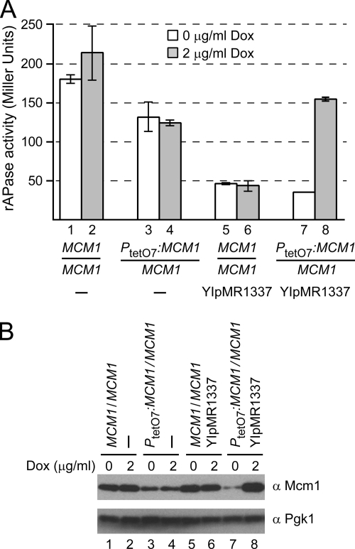
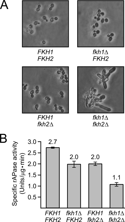
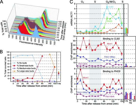
Cells devote considerable resources to nutrient homeostasis, involving nutrient surveillance, acquisition, and storage at physiologically relevant concentrations. Many Saccharomyces cerevisiae transcripts coding for proteins with nutrient uptake functions exhibit peak periodic accumulation during M phase, indicating that an important aspect of nutrient homeostasis involves transcriptional regulation. Inorganic phosphate is a central macronutrient that we have previously shown oscillates inversely with mitotic activation of PHO5. The mechanism of this periodic cell cycle expression remains unknown. To date, only two sequence-specific activators, Pho4 and Pho2, were known to induce PHO5 transcription. We provide here evidence that Mcm1, a MADS-box protein, is essential for PHO5 mitotic activation. In addition, we found that cells simultaneously lacking the forkhead proteins, Fkh1 and Fkh2, exhibited a 2.5-fold decrease in PHO5 expression. The Mcm1-Fkh2 complex, first shown to transactivate genes within the CLB2 cluster that drive G(2)/M progression, also associated directly at the PHO5 promoter in a cell cycle-dependent manner in chromatin immunoprecipitation assays. Sds3, a component specific to the Rpd3L histone deacetylase complex, was also recruited to PHO5 in G(1). These findings provide (i) further mechanistic insight into PHO5 mitotic activation, (ii) demonstrate that Mcm1-Fkh2 can function combinatorially with other activators to yield late M/G(1) induction, and (iii) couple the mitotic cell cycle progression machinery to cellular phosphate homeostasis.


Figures

FIG. 1.
FIG. 1.
PHO5 contains strong candidate Mcm1 and Fkh binding sites. (A) Alignment of Mcm1 and Fkh binding sites in the PHO5 promoter with those of prototypical cell cycle-regulated genes using CLUSTAL W (40). Residues highlighted with dark gray are fully conserved, whereas those highlighted with light gray occur in at least three of the five shown sequences. The lines above the consensus sequence (81) demarcate the core binding region for each factor. Nucleotides mutated (mut) in the PHO5 promoter in various strains are indicated in lowercase at the bottom. (B) Schematic representation of the PHO5 promoter. The putative Mcm1 binding site localizes to the linker between positioned nucleosomes −2 and −1, whereas the Fkh site is located in the edge of nucleosome −1 proximal to the TATA box. The bent arrow and downstream filled polygon designate the major transcription start site and coding region, respectively. UASp1 and UASp2, upstream-activating sequences p1 and p2 that associate with Pho4-Pho2.
FIG. 2.
FIG. 2.
Mcm1 protein levels are rate limiting for PHO5 mitotic activation. (A and B) Total rAPase activities (A) and immunoblotting (B) of Mcm1 (upper panel) and Pgk1 (lower panel) in diploid, WT MCM1/MCM1 (strain CCY694; bars 1 and 2), heterozygous PtetO7:MCM1/MCM1 (DNY2572; bars 3 and 4), heterozygous for tet-on regulators encoded on integrated YIpMR1337 (SPY3999; bars 5 and 6) and heterozygous for both PtetO7:MCM1 and YIpMR1337 (SPY4002, i.e., the experimental tet-on strain; bars 7 and 8) were assayed at mid-logarithmic phase in asynchronously growing YPD cultures treated with 0 or 2 μg of Dox/ml for 7 h in panel A (n = 2, mean ± range) or 16 h in panel B. The data in panel A are representative of six separate experiments; however, the results of a single experiment with duplicate cultures are shown since baseline rAPase activities vary between experiments. Diploid strains were used because MCM1 is an essential gene. The cellular morphology was unchanged in all strains, allowing normalization of activity levels to the OD600 of each culture. The error bars on bar 7 are too small to be visible. In an independent immunoblotting experiment (data not shown), a 6.5-h treatment of tet-on strain SPY4002 with 2 μg of Dox/ml also led to overexpression of Mcm1 compared to WT cells (strain CCY694).
FIG. 3.
FIG. 3.
Mcm1 is required for mitotic activation of PHO5. Haploid WT (MCM1 PHM4; strain CCY899, bars 1 and 2), tet-off MCM1 (PtetO7:MCM1 PHM4; MRY3665; bars 3 and 4), MCM1 phm4Δ (DNY2467; bars 5 and 6), and tet-off MCM1 phm4Δ (PtetO7:MCM1 phm4Δ; SPY3900; bars 7 and 8) cells were grown asynchronously to early logarithmic phase in YPD and then treated overnight as indicated with 0, 2, or 5 μg of Dox/ml (n = 2, mean ± range; the results are representative of three independent experiments). Cells were then viewed by light microscopy (A), assayed by immunoblotting with anti-Mcm1 (upper panel) or Pgk1 (lower panel) antibody (B), or assayed for total rAPase activity (C). Haploid phm4Δ strains (DNY2467 and SPY3900) were used to gauge the effects of loss of polyP on rAPase levels in the absence and presence of Dox. Since tet-off MCM1 cells adopt a filamentous morphology, specific rAPase activities of total cellular protein extracts were determined.
FIG. 4.
FIG. 4.
Deletion of FKH genes decreases rAPase activity. Haploid WT (FKH1 FKH2, strain SPY3718), single mutant (fkh1Δ FKH2, SPY3717; and FKH1 fkh2Δ, SPY3719), and double mutant (fkh1Δ fkh2Δ, SPY3716) strains were dissected from a single tetrad and then grown asynchronously to early logarithmic phase in YPD and visualized by light microscopy (A) or assayed for total rAPase activity (B). The specific rAPase activity of total protein extracts was determined in panel B because fkh1Δ fkh2Δ cells adopt a filamentous morphology (n = 2, mean ± range). The data are representative of two independent experiments.
FIG. 5.
FIG. 5.
Mitotic blockage per se does not lead to loss of PHO5 expression. Haploid WT CDC20 (DY2765) strains and PGAL1:CDC20 (DY6669) were cultured as described in Materials and Methods. Briefly, strains were grown in galactose-containing, defined Pi-free medium with Pi added back. Cells were then transferred after a washing step to glucose-containing, defined Pi-free medium with Pi added back for 3 h to repress PGAL1:CDC20 expression and effect M-phase arrest of strain DY6669, whereas strain DY2765 continued to grow asynchronously. After the 3-h arrest period, the cells were washed and transferred to the same medium containing either 0 or 13.4 mM Pi for 6 h before being assayed for rAPase activity.
FIG. 6.
FIG. 6.
Mutations in the candidate Mcm1 and Fkh binding sites of the PHO5 promoter impair mitotic activation. (A) rAPase activity of haploid WT (PPHO5, CCY577), single mutant (PPHO5-mcm1, DNY2768; and PPHO5-fkh, DNY2757), and double mutant (PPHO5-mcm1 fkh, DNY2850) strains determined from asynchronously growing YPD cultures (n = 2, mean ± range; the results are representative of two independent experiments). The Mcm1 and Fkh binding sites in the native genomic copy of the PHO5 promoter were mutated either singly or doubly as indicated in Fig. 1A. (B) PHO5 transcript levels in WT (PPHO5 bar1Δ, DNY1061) and double mutant (PPHO5-mcm1 fkh bar1Δ, DNY2970) strains in total RNA isolated at the indicated times after synchronous release from α-factor arrest. The top panel shows the results of RNA blotting for PHO5 and TCM1 mRNAs, and the bottom panel shows the quantified blotting results, normalizing PHO5 to TCM1 transcript levels for both strains.
FIG. 7.
FIG. 7.
Mcm1 and Pho4 induce PHO5 by parallel, nonredundant pathways. To test the effects of all possible combinations of loss of Mcm1 and/or forkhead protein binding and Pho4 function on mitotic induction of PHO5, PHO4 was deleted in haploid WT (PPHO5, CCY577 [A1]), single mutant (PPHO5-fkh, DNY2757 [B1]; and PPHO5-mcm1, DNY2768 [C1]), and double mutant (PPHO5-mcm1 fkh, DNY2850 [D1]) strains. Respectively, three independent pho4Δ transformants—SPY4125 to SPY4127 (PPHO5 pho4Δ [A2 to A4]), SPY4128 to SPY4130 (PPHO5-fkh pho4Δ [B2 to B4]), SPY4131 to SPY4133 (PPHO5-mcm1 pho4Δ [C2 to C4]), and SPY4134 to SPY4136 (PPHO5-mcm1 fkh pho4Δ [D2 to D4])—were isolated and tested for rAPase activity by spotting an equivalent number of cells on a YPD plate. Another pho4Δ strain (SHY2560) where PHO4 was deleted in a different haploid strain is spotted in row E. The darkness of each spot of cells is proportional to its rAPase activity. The data are representative of two independent experiments.
FIG. 8.
FIG. 8.
Cell cycle stage-specific association of Mcm1 and Fkh proteins with the PHO5 promoter. Doubly tagged strain DY12872 (cdc28-13ts FKH1-6HA FKH2-18Myc) was synchronously released from G1 arrest (37°C) and, at the indicated times, aliquots of cells were fixed with ethanol and stained with Sytox dye for flow cytometric analysis (A), fixed with ethanol for determination of budding index (B), and cross-linked with formaldehyde for ChIP analysis (C, middle and bottom panels). (C, top panel) Total RNA was also isolated from nonfixed cells for qRT-PCR of the indicated transcripts. ChIP enrichment (n = 2, mean ± range) is normalized to a region on chromosome 1 with no known ORFs (52). In parallel, an asynchronously growing culture of an untagged control strain (DY12878; No tag) was harvested in logarithmic (Log) phase and processed for budding index in panel B as well as ChIP in panel C. Note that the budding indices for times 70 to 130 min were not scored because buds were just growing in size and exhibited no change in morphology. The IgG normal serum control for the anti-Mcm1 antibody is also shown in panel C (middle and bottom panels).
FIG. 9.
FIG. 9.
M-phase arrest enriches Mcm1 binding to the PHO5 promoter. Doubly tagged strain DY12872 (cdc28-13ts FKH1-6HA FKH2-18Myc) was either arrested in G1 at 37°C or arrested and then released into medium without (−) or with (+) 100 μM Noc at 25°C for 150 min. Aliquots of cells were then cross-linked with formaldehyde for ChIP analysis. ChIP signals (n = 2, mean ± range) were determined for PHO5 and a control locus (HCM1 ORF) and normalized to the signals for each locus in an asynchronous (asynchr.) culture of strain DY12872.
FIG. 10.
FIG. 10.
Sds3, a component of the Rpd3L histone deacetylase complex, associates with the PHO5 promoter in G1 phase. Strain DY12247 (PGAL1:CDC20 SDS3-13Myc) was synchronously released from arrest in late M phase and, at the indicated times, fixed with formaldehyde for ChIP analysis. ChIP enrichment (n = 2, mean ± range) is normalized to an ORF-free region on chromosome 1 (52).
FIG. 11.
FIG. 11.
Genetic model for cell cycle-dependent regulation of PHO5. Pho4-Pho2 and Mcm1 induce the PHO5 promoter in G2/M through separate, nonredundant pathways. In a minor pathway, Mcm1 acts in conjunction with Fkh2 to activate PHO5 mitotic induction, possibly through recruitment of the Ndd1 coactivator (19, 34, 66). In addition, Mcm1 also appears to activate PHO5 via a pathway separate from Mcm1-Fkh2-Ndd1. This is suggested by the observation that secondary loss of polyP reserves and hence intracellular Pi depletion suppresses defects in PHO5 mitotic activation in cells deleted for both FKH genes but has no effect on cells lacking detectable Mcm1 protein. In this additional pathway, the question mark indicates that it is unclear whether Mcm1 exerts its action by itself, via interaction with an additional DNA-binding cofactor, through recruitment of a different coactivator, or some combination of these mechanisms. In G1 phase, Mcm1-Fkh2 may act as a repressor through recruitment of the Rpd3L histone deacetylase complex (Fig. 10), probably through Fkh2 (87, 92). It is currently unknown whether this recruitment is direct or indirect.

References

    1. Althoefer, H., A. Schleiffer, K. Wassmann, A. Nordheim, and G. Ammerer. 1995. Mcm1 is required to coordinate G2-specific transcription in Saccharomyces cerevisiae. Mol. Cell. Biol. 155917-5928. - PMC - PubMed
    1. Amon, A., M. Tyers, B. Futcher, and K. Nasmyth. 1993. Mechanisms that help the yeast cell cycle clock tick: G2 cyclins transcriptionally activate G2 cyclins and repress G1 cyclins. Cell 74993-1007. - PubMed
    1. Anderson, M. S., and J. M. Lopes. 1996. Carbon source regulation of PIS1 gene expression in Saccharomyces cerevisiae involves the MCM1 gene and the two-component regulatory gene, SLN1. J. Biol. Chem. 27126596-26601. - PubMed
    1. Barbaric, S., T. Luckenbach, A. Schmid, D. Blaschke, W. Hörz, and P. Korber. 2007. Redundancy of chromatin remodeling pathways for the induction of the yeast PHO5 promoter in vivo. J. Biol. Chem. 28227610-27621. - PubMed
    1. Barbaric, S., M. Münsterkötter, C. Goding, and W. Hörz. 1998. Cooperative Pho2-Pho4 interactions at the PHO5 promoter are critical for binding of Pho4 to UASp1 and for efficient transactivation by Pho4 at UASp2. Mol. Cell. Biol. 182629-2639. - PMC - PubMed

Publication types

MeSH terms