. 2009 Aug 1;122(Pt 15):2716-26.
doi: 10.1242/jcs.043794. Epub 2009 Jul 13.

Nesprin-2 interacts with meckelin and mediates ciliogenesis via remodelling of the actin cytoskeleton

Affiliations

Nesprin-2 interacts with meckelin and mediates ciliogenesis via remodelling of the actin cytoskeleton

Helen R Dawe et al. J Cell Sci. .

Abstract

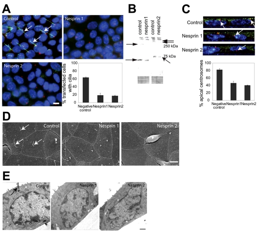
Meckel-Gruber syndrome (MKS) is a severe autosomal recessively inherited disorder caused by mutations in genes that encode components of the primary cilium and basal body. Here we show that two MKS proteins, MKS1 and meckelin, that are required for centrosome migration and ciliogenesis interact with actin-binding isoforms of nesprin-2 (nuclear envelope spectrin repeat protein 2, also known as Syne-2 and NUANCE). Nesprins are important scaffold proteins for maintenance of the actin cytoskeleton, nuclear positioning and nuclear-envelope architecture. However, in ciliated-cell models, meckelin and nesprin-2 isoforms colocalized at filopodia prior to the establishment of cell polarity and ciliogenesis. Loss of nesprin-2 and nesprin-1 shows that both mediate centrosome migration and are then essential for ciliogenesis, but do not otherwise affect apical-basal polarity. Loss of meckelin (by siRNA and in a patient cell-line) caused a dramatic remodelling of the actin cytoskeleton, aberrant localization of nesprin-2 isoforms to actin stress-fibres and activation of RhoA signalling. These findings further highlight the important roles of the nesprins during cellular and developmental processes, particularly in general organelle positioning, and suggest that a mechanistic link between centrosome positioning, cell polarity and the actin cytoskeleton is required for centrosomal migration and is essential for early ciliogenesis.


Figures

Fig. 1.
Fig. 1.
Domain structures, isoform diversity and subcellular localization of meckelin and nesprin-2. (A) Domain structure of meckelin showing signal peptide, cysteine-rich-repeat region, three transmembrane domains and a coiled-coil domain. The location of the epitope for anti-meckelin antibody R29 is indicated, as is the yeast two-hybrid (Y2H) bait fragment. (B) Domain structure of the GIANT (NUANCE) isoform of nesprin-2 showing the ABD, SMC domain, spectrin-repeat regions and a transmembrane KASH domain. The locations for the epitopes of the anti-nesprin-2 antibodies K20-478 (ABD) and K49-260 (spectrin repeats) are indicated. Numbers represent amino acid residues. (C) Immunoblotting of IMCD3 WCE at 48 hours prior to confluence (–48), confluence (0) and 72 hours post-confluence (+72) with anti-meckelin antibody (R29) demonstrated that throughout ciliogenesis (–48 to +72 hours) full-length meckelin (120 kDa) was present. Smaller species of meckelin (80, 60 and 55 kDa) were also present at subconfluence and confluence. Immunoblotting with anti-nesprin-2 antibody K20-478 showed high levels of a 250-kDa ABD-containing isoform at post-confluence only, with a second major ABD-containing isoform of 55 kDa present before and after confluence. A minor 150-kDa isoform was also seen at confluence and post-confluence. Immunoblotting with anti-nesprin-2 antibody K49-260 demonstrated that a large (>350 kDa) spectrin-repeat-containing isoform persisted throughout ciliogenesis. A 250-kDa spectrin-repeat-containing isoform was present at moderate levels at subconfluence only, but other smaller species were present at all time points. An immunoblot for a loading control (β-actin) was used for each time point. (D) Co-immunostaining and confocal microscopy of ciliated IMCD3 cells at post-confluence (+72 hours) with an anti-meckelin antibody (R29; first column) and an anti-acetylated-α-tubulin antibody (second column). A merged image shows meckelin in green, acetylated-α-tubulin in red and a DAPI-labelled nucleus in blue. The upper panels show a 0.1-μm section above the apical surface of the cell showing a punctate localization of meckelin at the ciliary membrane. The middle panels are a whole-cell projection showing meckelin at the basal body (arrowhead). The bottom panels show a confocal xz projection through the cell monolayer, from the apical to basal surfaces. (E) Immunostaining and confocal microscopy of post-confluent (+72 hours) ciliated IMCD3 cells with anti-nesprin-2 antibody K49-260 demonstrated that spectrin-repeat-containing isoforms of nesprin-2 (red) have a perinuclear localization. The nucleus was visualized with DAPI. Scale bars: 5 μm.
Fig. 2.
Fig. 2.
Meckelin interacts with nesprin-2. (A) Immunoblotting of WCE from HA-MKS3-transfected (TF) HEK 293 cells confirmed expression of HA-tagged meckelin, at approximately 130 kDa. Immunoprecipitation (IP) of the same WCE with anti-nesprin-2 antibody against the ABD (K20-478) confirmed an interaction with HA-tagged meckelin. (B) Pulldown of ABD-containing isoforms of nesprin-2 from subconfluent HEK 293 WCE by GST-tagged meckelin C-terminus (Ct) but not by GST alone. Immunoblotting was performed using antibody K20-478. Arrows indicate 150- and 55-kDa ABD-containing isoforms of nesprin-2. HEK293 indicates 10% of total input WCE. (C) IP of ABD-containing nesprin-2 isoforms (arrows indicate 150-kDa and 250-kDa isoforms) from confluent HEK 293 WCE with anti-meckelin (R29) and anti-MKS1 but not by a no-antibody control, pre-immune serum negative control or two irrelevant antibodies (anti-parkin and anti-EFe4). HEK293 indicates 5% of total input WCE, and the asterisk (*) indicates the position of IgG heavy chain (size approximately 55 kDa). (D) IP of endogenous meckelin (arrow, size 120 kDa) from HEK 293 WCE with anti-nesprin-2 (K20-478), but not by a no-antibody control or two irrelevant antibodies. (E) IP of MKS1 (arrow, size 70 kDa) from HEK 293 WCE with anti-nesprin-2 (K20-478), but not by either a no-antibody control or the two irrelevant antibodies. (F) Subcellular localization of meckelin and nesprin-2. Co-immunostaining and confocal microscopy of IMCD3 cells at subconfluence (–48 and –24), confluence (0) and post-confluence (+24) with affinity-purified anti-meckelin antibody (green channel) and anti-nesprin-2 antibody against the ABD (K20-478, red channel). At –48 hours, a meckelin–nesprin-2 colocalization was seen at the cell periphery at probable filopodia or microspikes (arrowheads; inset shows 3× enlargement) that is absent at later time points. At –24 hours, a meckelin tubulovesicular pattern was seen that partially colocalized with nesprin-2 tubulovesicular elements (arrows). This meckelin pattern persisted, but the nesprin-2 tubulovesicular pattern was absent at confluence and post-confluence. Nesprin-2 persisted at the nucleus throughout, but perinuclear localization (arrowhead) occurred only in post-confluent ciliated cells (+24). Scale bar: 5 μm.
Fig. 3.
Fig. 3.
Loss of nesprins prevents ciliogenesis and causes defects in nuclear morphology, but does not affect apical-basal polarity or nuclear positioning. (A) Immunofluorescence of IMCD3 monolayers transfected with negative-control siRNA, siRNA against nesprin-1 or siRNA against nesprin-2. Note the loss of primary cilia (acetylated α-tubulin staining, green) in nesprin-1 (Syne1)- and nesprin-2 (Syne2)-silenced cells compared with the negative control. DNA is labelled with DAPI (blue) and the centrosome (γ-tubulin staining) is shown in red. The graph shows quantification of the presence of a primary cilium in transfected cells, expressed as mean ± s.e.m. of three independent experiments. At least 1500 cells were counted for each condition. Scale bar: 5 μm. (B) Western blots to show the success of RNAi. Immunoblots showing certain isoforms of nesprin-1 (left panels; indicated by arrows) and isoforms of nesprin-2 that contain the ABD (right panels; arrows) are partially depleted after 96 hours of siRNA treatment, consistent with a transfection efficiency of ∼60%, compared with the negative-control-transfected lane. Ponceau stain of immunoblots is shown as a loading control. (C) Confocal xz projections through the cell monolayer of cells (from the apical to basal surfaces); cells were transfected with negative-control siRNA (top panel), siRNA against nesprin-1 (middle panel) or siRNA against nesprin-2 (bottom panel). Cells were immunostained with anti-moesin to mark the apical cell surface (green), DAPI for DNA (blue) and anti-γ-tubulin for centrosomes (red). Note the centrosome (arrows) is deep inside the cell in nesprin-1 (Syne1)- and nesprin-2 (Syne2)-silenced cells, compared with its apical position above the nucleus in control cells. The graph shows the quantification of centrosome positioning expressed as mean ± s.e.m. of three independent experiments. At least 700 cells were counted per condition. `Apical' is defined as the most apical 1.5-μm region of the cell. (D) Scanning electron microscopy of IMCD3 monolayers transfected with negative-control siRNA, siRNA against nesprin-1 or siRNA against nesprin-2. Microvilli form on the apical surface despite the lack of cilia. Arrows indicate primary cilia in control cells. Scale bar: 5 μm. (E) Transmission electron micrographs of control, nesprin-1 (Syne1)-silenced and nesprin-2 (Syne2)-silenced cells showing that the distance between the top of the nucleus and the apical cell surface is not affected by loss of nesprin-1 or nesprin-2. Note the primary cilium (arrow) in the control panel, and the invaginated nuclei observed on loss of nesprin-1 and especially nesprin-2. Scale bar: 1 μm.
Fig. 4.
Fig. 4.
Subcellular phenotype of meckelin loss. (A) siRNA knockdown of Mks3 in IMCD3 cells. The top panels show IMCD3 cells co-immunostained with anti-γ-tubulin antibody (green) and an anti-acetylated-α-tubulin antibody (red). In cells transfected with scrambled siRNA control (scr.), cilia are intact, but in cells transfected with Mks3 siRNA (Mks3), cilia are absent. DAPI staining of nuclei is shown in blue. Bottom panels show immunoblotting of WCE from cells transfected with scrambled siRNA and Mks3 siRNA, showing loss of meckelin in the latter compared with a loading control (β-actin). Scale bars: 5 μm. (B) Subconfluent (–48 hours) IMCD3 cells co-immunostained with anti-meckelin (green), anti-nesprin-2 (K20-478; red) and phalloidin-labelled F-actin (blue). In cells transfected with scrambled siRNA control (scr.), nesprin-2 has a perinuclear distribution and meckelin a characteristic tubulovesicular pattern. In cells transfected with Mks3 siRNA, nesprin-2 colocalized with actin stress fibres at the cell periphery and within the cytoplasm (arrowheads). Scale bars: 5 μm. (C) Subcellular phenotypes of immortalized human embryonic fibroblasts cultured from undiseased controls (con. 1 and con. 2) and an individual with Meckel-Gruber syndrome (MKS), compound heterozygous for the missense and truncating mutations [p.M261T]+[p.R217X] in MKS3. The top panels show control fibroblasts (con. 1) and MKS fibroblasts co-immunostained for γ-tubulin (green) and acetylated-α-tubulin (red). In control fibroblasts, cilia are intact, but in MKS3-mutated fibroblasts (MKS) cilia are absent. DAPI staining of nuclei is shown in blue. Immunoblots with anti-meckelin antibody and a loading control (β-actin) on WCE from two control fibroblast cell lines and MKS fibroblasts confirm a significant reduction in overall meckelin levels in MKS3-mutated fibroblasts compared with controls. The graph shows a cilia count in which ten high-power fields were analyzed for the presence of cilia, and confirmed significant cilia loss from MKS fibroblasts (P<0.001) compared with controls. In the scatter plot, the position of individual centrosomes (red dots) in multiple cells (n=100) is displayed relative to the edge of the nucleus. Centrosomes in control cells lie within <0.5 μm of the nucleus, whereas those in MKS fibroblasts are on average 2 μm from the nucleus (P<0.001; mean values are shown by black lines and s.d. in grey). Scale bars: 5 μm. (D) Subconfluent (–48hr) fibroblasts and post-confluent fibroblasts (+48hr) co-immunostained for meckelin (green), ABD-containing nesprin-2 (K20-478; red) and phalloidin (blue). In control fibroblasts (con.), nesprin-2 has a characteristic perinuclear distribution. In MKS fibroblasts, nesprin-2 also colocalized with actin stress fibres at the cell periphery and within the cytoplasm (arrowheads). This localization of nesprin-2 persists at sub- and post-confluent time points. In subconfluent (–48hr) fibroblasts, meckelin has a perinuclear and tubulovesicular pattern in control cells, and colocalizes with nesprin-2 at actin-rich filopodia or microspikes. This distribution of meckelin is absent in MKS3-mutated cells. Arrowheads indicate stress fibres. Scale bars: 5 μm. (E) RhoA-activation assay of normal control and MKS fibroblast WCEs showed a 4.8-fold increase in the levels of activated RhoA-GTP in patient cells. Total RhoA and β-actin are shown as loading controls. A positive control for the assay (+; loading with non-hydrolyzable GTPγS) and a negative control (–; loading with GDP) are also shown. (F) Alterations in nuclear morphology with loss of meckelin. Nuclei were labelled with DAPI. In IMCD3 cells transfected with scrambled siRNA control (scr.) and in control fibroblasts (Fibs), nuclear shape was uniformly oval (first column). In IMCD3 cells transfected with Mks3 siRNA and in MKS fibroblasts a proportion of nuclei were abnormal, with nuclear invaginations and nuclear blebbing (arrowheads). A proportion of abnormal MKS fibroblast nuclei also contained vacuoles (arrow). Scale bars: 5 μm. The graph quantifies the proportions of abnormal nuclei in fibroblasts and in siRNA-transfected IMCD3 cells. MKS fibroblasts have significantly more abnormal nuclei (P<0.001), as do IMCD3 cells transfected with Mks3 siRNA (P<0.01).
Fig. 5.
Fig. 5.
Model of the interactions of MKS proteins with nesprin-2 isoforms, and their possible effects on ciliogenesis. Confirmed biochemical interactions between the coiled-coil domain of meckelin, MKS1 and nesprin-2 are shown as red arrows. Meckelin might also mediate ROCK-RhoA signalling (black arrows) to regulate the activity of myosin II and interactions with the actin cytoskeleton (pale-blue rods). Actin-cytoskeleton remodelling mediated by RhoA, and enrichment at the apical cell surface, could allow docking of the centrosome/basal body (green arrow). MKS1 (pale green) is a known component of the basal body, and interacts with both meckelin and actin-binding isoforms of nesprin-2. The domain structure of nesprin-2 is indicated (refer to Fig. 1B). Because KASH-domain-containing isoforms of nesprin-2 are also known to mediate nuclear repositioning, these isoforms could also mediate microtubule-dependent centrosome migration to the apical cell surface (grey arrow). Numbers refer to amino acid residues. PM, plasma membrane; ONM, outer nuclear membrane; PS, perinuclear space; INM, inner nuclear membrane.

References

    1. Adams, M., Smith, U. M., Logan, C. V. and Johnson, C. A. (2008). Recent advances in the molecular pathology, cell biology and genetics of ciliopathies. J. Med. Genet. 45, 257-267. - PubMed
    1. Baala, L., Romano, S., Khaddour, R., Saunier, S., Smith, U. M., Audollent, S., Ozilou, C., Faivre, L., Laurent, N., Foliguet, B. et al. (2007). The Meckel-Gruber syndrome gene, MKS3, is mutated in Joubert syndrome. Am. J. Hum. Genet. 80, 186-194. - PMC - PubMed
    1. Boisvieux-Ulrich, E., Laine, M. and Sandoz, D. (1990). Cytochalasin D inhibits basal body migration and ciliary elongation in quail oviduct epithelium. Cell Tissue Res. 259, 443-454. - PubMed
    1. Burakov, A., Nadezhdina, E., Slepchenko, B. and Rodionov, V. (2003). Centrosome positioning in interphase cells. J. Cell Biol. 162, 963-969. - PMC - PubMed
    1. Chevrier, V., Piel, M., Collomb, N., Saoudi, Y., Frank, R., Paintrand, M., Narumiya, S., Bornens, M. and Job, D. (2002). The Rho-associated protein kinase p160ROCK is required for centrosome positioning. J. Cell Biol. 157, 807-817. - PMC - PubMed

Publication types

MeSH terms