. 2009 Sep;37(16):5511-28.
doi: 10.1093/nar/gkp571. Epub 2009 Jul 13.

The role of deadenylation in the degradation of unstable mRNAs in trypanosomes

Affiliations

The role of deadenylation in the degradation of unstable mRNAs in trypanosomes

Angela Schwede et al. Nucleic Acids Res. 2009 Sep.

Abstract

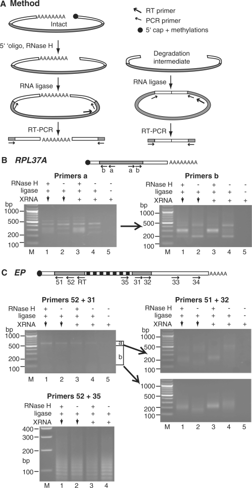
Removal of the poly(A) tail is the first step in the degradation of many eukaryotic mRNAs. In metazoans and yeast, the Ccr4/Caf1/Not complex has the predominant deadenylase activity, while the Pan2/Pan3 complex may trim poly(A) tails to the correct size, or initiate deadenylation. In trypanosomes, turnover of several constitutively-expressed or long-lived mRNAs is not affected by depletion of the 5'-3' exoribonuclease XRNA, but is almost completely inhibited by depletion of the deadenylase CAF1. In contrast, two highly unstable mRNAs, encoding EP procyclin and a phosphoglycerate kinase, PGKB, accumulate when XRNA levels are reduced. We here show that degradation of EP mRNA was partially inhibited after CAF1 depletion. RNAi-targeting trypanosome PAN2 had a mild effect on global deadenylation, and on degradation of a few mRNAs including EP. By amplifying and sequencing degradation intermediates, we demonstrated that a reduction in XRNA had no effect on degradation of a stable mRNA encoding a ribosomal protein, but caused accumulation of EP mRNA fragments that had lost substantial portions of the 5' and 3' ends. The results support a model in which trypanosome mRNAs can be degraded by at least two different, partially independent, cytoplasmic degradation pathways attacking both ends of the mRNA.


Figures

Figure 1.
Figure 1.
Cartoon maps of PAN2 and PAN3 from various species, as indicated on the left. Domains recognized by Prosite are also shown. Sc: Saccharomyces cerevisiae; Hs: Homo sapiens; Tb: T. brucei; Clamy: Chlamydomonas reinhardtii; Thp: Thalassiosira pseudonana; Neig: Naegleria fowleri; Aure: Aureococcus anophagefferens; Micp: Micromonas pusilla. EXOc is the exoribonuclease domain; ‘Kin’ is the putative kinase domain. The downward arrows indicate the approximate positions of residues thought to be required for catalytic activity according to an NCBI domain search and specifications for the cd06143 sequence cluster [e.g. (114,115)]. Complete sequence alignments are provided as Figures S1 (PAN2) and S2 (PAN3).
Figure 2.
Figure 2.
PAN2 is mainly in the cytoplasm and RNAi inhibits trypanosome growth. (A) In bloodstream-form or procyclic-form trypanosomes, one PAN2 allele was tagged in situ at the N terminus with a sequence encoding a V5 tag. The tag was detected by immunofluorescence. DAPI staining shows the positions of the nucleus (large spot) and kinetoplast DNA (small dot). A phase-contrast image of the trypanosome is on the right. (B) Detergent lysates (T) from the V5-tagged cells were separated into nuclear (N) and cytoplasmic (C) fractions and proteins detected by western blotting. XRND: nuclear exoribonuclease (61); TR: trypanothione reductase. (C) PAN2 RNAi inhibits trypanosome growth. Results for two bloodstream-form cell lines are shown; each expresses the tet repressor and T7 polymerase (1313-514) and has an integrated copy of the PAN2 RNAi plasmid. Cells were grown with (open circles, dashed lines) or without (closed circles) tetracycline added at day 0, and were diluted as required. Cumulative growth curves are shown, with doubling times calculated from exponential curves (Kaleidograph). The results are for two lines expressing the CAT-GC-EP reporter. The panels beneath are northern blots probed for PAN2, with 7SL as a control. In the right hand panel, PAN2 mRNA was only depleted by 30%, but the cells nevertheless showed growth inhibition. (D) RNAi in cells with V5 in situ tagged PAN2, details as in (C). The panel below is a western blot that was probed with antibody to the V5 tag and to aldolase.
Figure 3.
Figure 3.
PAN2 depletion inhibits deadenylation. (A) Trypanosome RNAs were 3′ labelled with [32P]-pCp then digested with RNase T1 and RNase A to leave the poly(A) tails. The resulting RNA was separated on a denaturing polyacrylamide gel and the [32P] was detected using a phosphorimager. The downward arrows indicate depletion of PAN2 by RNAi. The ‘+’ sign indicates that PAN2 is present (RNAi line in the absence of tetracycline). (B) Quantitation of (A). The poly(A) tails were divided into three size classes, as indicated on the right, and the signal from each class measured. The proportion of each class, relative to the total signal on the relevant lane, was then measured. This quantitation minimizes the effect of PAN2 depletion since it does not show the differences in total signal intensity between lanes. Results are the arithmetic mean of three independent measurements with standard deviations. Individual sets of three measurements that were significantly different in a Student's t-test (P < 0.05, 30-min time points) are shown with asterisks. The results for 5 min, 15 min and 30 min were also pooled and compared using a Wilcoxon ranking test (see numbers above the lanes). Tails of 0–50 nt were less abundant in the PAN2 RNAi samples than in the control in 15/15 cases, giving a P-value of <0.01. The longer tails of 51–100 nt and 101–200 nt were more abundant in the PAN2 RNAi samples (P-values of 0.02 and <0.01, respectively). (C) Northern blots showing amounts of various mRNAs after inhibition of mRNA synthesis. Sinefungin was added at time –5 min, and Actinomycin D was added at time = 0. ACT: actin; HISH4: Histone H4; 7SL: RNA of the signal recognition particle. Lanes labelled A- contain RNA that was incubated with oligo d(T) and RNase H before loading. Signals were detected and measured using a phosphorimager, using the 7SL RNA as a loading control. (D) Quantitation of results for the HISH4 mRNA. Means and standard deviations for four independent measurements are shown, with fitted exponential degradation curves (Kaleidograph) and corresponding half-lives. For quantitation of all measurements relative to wild-type values see Figure S3. (E) Quantitation of results for the ACT mRNA. Means and standard deviations for five independent measurements are shown. The result of a Student's t-test for the 30-min time point is indicated on the graph. The differences at 30 min were also significant (P < 0.02) in the Wilcoxon ranking test.
Figure 4.
Figure 4.
Degradation of trypanosome EP1 mRNA is effected by deadenylation-dependent and -independent pathways. (A) mRNA synthesis was inhibited with Sinefungin and Actinomycin D in cells with inducible CAF1 RNAi, either without tetracycline or 18 h after tetracycline addition. Total RNA was prepared and the abundances of specific transcripts analysed by northern blotting. A typical blot is shown. Quantitation is on the right. Results are arithmetic mean and standard deviation for four independent experiments. The fitted lines were determined by assuming the presence of two exponential decay components and minimising deviation from the experimental observations (see Methods section). The parameters used to fit the curves are indicated as half-life in min, and the proportion of the RNA with that half-life. (B) As for A, but using cells with RNAi targeting PAN2. Results are for three experiments, and P-values from a Student's t-test for the 30 min and 60 min time points are shown. (C) Degradation kinetics of EP mRNA in cells with normal or depleted XRNA. Data points for two independent experiments are shown, with a different symbol for each experiment. For the wild-type (no integrated RNAi construct), one experiment was included as a control. The curves are fitted to the arithmetic mean values.
Figure 5.
Figure 5.
Depletion of CAF1 or PAN2 has minor effects on degradation of CAT-PGKB mRNA. (A) Northern blot illustrating CAT-PGKB mRNA degradation in cells depleted of CAF1. Details as in Figure 4A, three experiments. (B) Northern blot illustrating CAT-PGKB mRNA degradation in cells depleted of PAN2. Details as in Figure 4B.
Figure 6.
Figure 6.
Degradation of the CAT-GC-EP reporter mRNA in trypanosomes. (A) Possible pathways of degradation. The CAT-GC-EP mRNA has, from 5′ to 3′, a cap (small circle), a short 5′UTR (white fill), a CAT open reading frame (black), a G30C30 sequence, then the EP1 3′-UTR (white, with destabilising 26-mer in grey) and poly(A) tail (AAAA). (1) deadenylation as the first step in degradation (product ‘A’); (2) degradation of the deadenylated RNA from the 3′-end, pausing at the G30C30 sequence to produce the CAT-GC intermediate (product ‘B’); (3) decapping of the deadenylated mRNA; product ‘C’, indistinguishable from ‘A’ by northern blotting; (4) degradation of product ‘C’ from the 5′-end, pausing at the G30C30 sequence to give the deadenylated 3′ GC-EP RNA (product ‘E’); (5) decapping as the first step in degradation, to give product ‘F’, indistinguishable from the initial RNA by northern blotting; (6) degradation of product ‘F’ from the 5′-end, pausing at G30C30 to give a polyadenylated 3′ GC-EP RNA (product ‘D’); (7) deadenylation of RNA ‘D’ to give RNA ‘E’. (B) The effect of inducible CAF1 RNAi on CAT-GC-EP mRNA degradation. CAF1+: cells without tetracycline; CAF1 with downward arrow: cells with tetracycline (19–21.5 h). Total RNA (T) was prepared and part of it was separated into poly(A)+ (A+) and poly(A)– (A–) fractions. The identities of the bands are shown on the left and sizes on the right. The upper panels were hybridized with an EP 3′-UTR riboprobe. This reproducibly cross-hybridizes with the smallest rRNA fragment. Although this signal has been cropped from the picture, it smears a little into the upper portions of the total and poly(A)– lanes. The lower panels are hybridizations with a CAT probe. The ratio of the CAT-GC fragment to the full-length mRNA fragment was measured in three experiments: for cells without tetracycline the average ratio was 21% (range 15–26%) and for CAF1-depleted cells 14% (range 12–16%). The degradation fragments were detected readily only if we probed for them first: the upper and lower panels therefore originate from different experiments.
Figure 7.
Figure 7.
RT–PCR of circularized mRNAs. (A) Illustration of the method for intact, capped RNA (right) and a degradation intermediate (left). The open reading frame is filled grey, other symbols are shown on the figure. For detailed description see text. (B) Ethidium-bromide stained gels of amplified products for RPL37A. A cartoon of the mRNA, not to scale, showing the positions of the primers, is above the gels. Products from the first PCR (primers ‘a’) are shown on the left, and from the second, nested PCR (primers ‘b’) on the right. (C) Ethidium-bromide stained gels of amplified products for EP. In the cartoon, the striped region represents the segment encoding EP repeats. After the first PCR with primers 52 and 31, DNA was isolated from the upper and lower portions of the gel, as indicated, and used for the nested PCR using primers 51 and 32. A separate picture shows the ladders obtained using primers 35 and 52. In each case, the area of the gel containing most DNA was excised for purification and cloning of the PCR products, and clones were selected randomly for sequencing. Gels for amplifications with 3′ primers 33 or 34, with 5′ primer 51, looked very similar to the lower panel for primers 51 and 32; the same procedure was followed but only clones with the longest inserts were sequenced.
Figure 8.
Figure 8.
(A) RNAi targeting T. brucei MEX67 inhibits growth. A cumulative growth curve is shown. (B and C) RNAi targeting T. brucei MEX67 results in accumulation of poly(A)+ RNA in the nucleus. Cells without an RNAi construct (B) or cells with inducible RNAi (C) were incubated with or without tetracycline for 48 h. poly(A) was detected in the cells by in situ hybridization (p(A)). The DNA stain (DAPI) is on the right; some outlines of the parasites are added (dotted lines) to facilitate interpretation. (D) Effect of MEX67 RNAi on mRNA levels. RNA was prepared at the indicated times after tetracycline addition and EP, PGK, and 7SL mRNAs were detected. (E) Quantitation of two experiments as in (D). The amount of PGKC mRNA in cells without an RNAi construct (WT) was set to 1.0; for EP and PGKB, the amount of mRNA at 48 h was set to 1.0. A third experiment gave similar results but the signals were too faint for quantitation.

Similar articles

Cited by

References

    1. Caponigro G, Parker R. Multiple functions for the poly(A)-binding protein in mRNA decapping and deadenylation in yeast. Genes Dev. 1995;9:2421–2432. - PubMed
    1. Tharun S, Parker R. Targeting an mRNA for decapping: displacement of translation factors and association of the Lsm1p-7p complex on deadenylated yeast mRNAs. Mol. Cell. 2001;8:1075–1083. - PubMed
    1. Wilusz CJ, Gao M, Jones CL, Wilusz J, Peltz SW. Poly(A)-binding proteins regulate both mRNA deadenylation and decapping in yeast cytoplasmic extracts. RNA. 2001;7:1416–1424. - PMC - PubMed
    1. Körner CG, Wormington M, Muckenthaler M, Schneider S, Dehlin E, Wahle E. The deadenylating nuclease (DAN) is involved in poly(A) tail removal during the meiotic maturation of Xenopus oocytes. EMBO J. 1998;17:5427–5437. - PMC - PubMed
    1. Dehlin E, Wormington M, Körner CG, Wahle E. Cap-dependent deadenylation of mRNA. EMBO J. 2000;19:1079–1086. - PMC - PubMed

Publication types

MeSH terms