. 2009 Oct;53(10):4200-10.
doi: 10.1128/AAC.00428-09. Epub 2009 Jul 13.

Staphylococcus aureus mutant screen reveals interaction of the human antimicrobial peptide dermcidin with membrane phospholipids

Affiliations

Staphylococcus aureus mutant screen reveals interaction of the human antimicrobial peptide dermcidin with membrane phospholipids

Min Li et al. Antimicrob Agents Chemother. 2009 Oct.

Abstract

Antimicrobial peptides (AMPs) form an important part of the innate host defense. In contrast to most AMPs, human dermcidin has an anionic net charge. To investigate whether bacteria have developed specific mechanisms of resistance to dermcidin, we screened for mutants of the leading human pathogen, Staphylococcus aureus, with altered resistance to dermcidin. To that end, we constructed a plasmid for use in mariner-based transposon mutagenesis and developed a high-throughput cell viability screening method based on luminescence. In a large screen, we did not find mutants with strongly increased susceptibility to dermcidin, indicating that S. aureus has no specific mechanism of resistance to this AMP. Furthermore, we detected a mutation in a gene of unknown function that resulted in significantly increased resistance to dermcidin. The mutant strain had an altered membrane phospholipid pattern and showed decreased binding of dermcidin to the bacterial surface, indicating that dermcidin interacts with membrane phospholipids. The mode of this interaction was direct, as shown by assays of dermcidin binding to phospholipid preparations, and specific, as the resistance to other AMPs was not affected. Our findings indicate that dermcidin has an exceptional value for the human innate host defense and lend support to the idea that it evolved to evade bacterial resistance mechanisms targeted at the cationic character of most AMPs. Moreover, they suggest that the antimicrobial activity of dermcidin is dependent on the interaction with the bacterial membrane and might thus assist with the determination of the yet unknown mode of action of this important human AMP.


Figures

FIG. 1.
FIG. 1.
Establishment of a bioluminescence-based screen for S. aureus mutants with altered resistance to dermcidin. (A) Relationship between bioluminescence count (RLU, relative light units) and the numbers of CFU; (B) comparison of OD and bioluminescence during 12 h of in vitro growth of S. aureus Xen36; (C) determination of the optimal dermcidin concentration to be used in the screen.
FIG. 2.
FIG. 2.
Dermcidin resistance of S. aureus Xen36 fecCD deletion mutant. The resistance to dermcidin of S. aureus Xen36 wild-type, fecCD deletion mutant, genetically complemented mutant (with plasmid pTfecCD), and control (with control plasmid pT181) strains was compared by bioluminescence assay (A) and conventional killing assays (B) by colony counting.
FIG. 3.
FIG. 3.
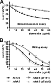
Dermcidin resistance of S. aureus Xen36 dak2 mutant. The resistance to dermcidin of S. aureus Xen36 wild-type, dak2 transposon mutant, genetically complemented mutant (with plasmid pTdak2), and control (with control plasmid pT181) strains was compared by bioluminescence assay (A) and conventional killing assays by colony counting (B).
FIG. 4.
FIG. 4.
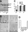
(A and B) TLC of S. aureus Xen36 dak2 mutant membrane phospholipids. The one-dimensional (A) and two-dimensional (B) TLCs of the dak2 mutant in comparison to those of the Xen36 wild-type and complemented mutant strains are shown. The left lanes in panel A and the top panel in panel B show the results for the PG and DPG standards. (C) Results of quantitative analysis of one-dimensional TLC for the three phospholipid species. ***, P < 0.001 (one-way ANOVA, Bonferroni posttest). (D) Cytochrome c binding assays. The degree of binding of the cationic protein cytochrome c to cells of the indicated strains was determined. There were no significant differences in the degrees of binding between the Xen36 wild-type strain, its isogenic dak2 mutant, and the complemented and control strains. The differential binding of cytochrome c to the S. aureus SA113 dlt mutant, which is defective in d-alanylation of teichoic acids, in comparison to that to wild-type strain SA113 was used as a positive control.
FIG. 5.
FIG. 5.
FAME analysis of the S. aureus Xen36 dak2 mutant membrane phospholipid fatty acids. FAME analysis by GC-MS was performed as described in Materials and Methods, and the resulting chromatograms were normalized for the highest peak (set equal to 100%) and superimposed. The main fatty acid species are labeled. Str, straight. Phospholipid preparations were from bacteria grown to stationary growth phase. Similar results were obtained from bacteria grown to mid-logarithmic growth phase, and the complemented mutant strain showed a pattern similar to that of the wild-type strain (data not shown).
FIG. 6.
FIG. 6.
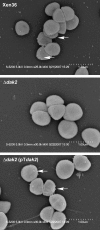
Immuno-SEM of S. aureus Xen36, dak2 mutant, and complemented mutant strains. Immuno-SEM pictures were obtained in the backscatter imaging mode after incubation with anti-dermcidin antisera (1:200) and subsequent incubation with gold-labeled anti-IgG. The arrows show examples of gold particles.
FIG. 7.
FIG. 7.
Specificity of dermcidin-phospholipid interaction. The levels of binding of dermcidin and cationic AMPs to phospholipid preparations from S. aureus Xen36 wild-type, dak2 mutant, complemented (with plasmid pTdak2), and control (with control plasmid pT181) strains are shown. Detection was performed by immuno-dot blotting with alkaline phosphatase-coupled second antibody. The results are from three independent experiments. Statistical analysis was by one-way ANOVA with Bonferroni posttests. All significant differences are marked, except for those against the results for PBS. *, P < 0.05; ***, P < 0.001.
FIG. 8.
FIG. 8.
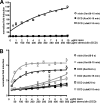
Assessment of pore-forming activity of dermcidin. To test whether dermcidin forms pores in the bacterial membrane, ATP release assays (A) and membrane depolarization assays with the membrane potential-sensitive fluorescent dye DiSC3(5) (B) were performed, and the results were compared to those obtained with the pore-forming peptide nisin. Values were taken at 15, 30, and 60 min (A) or 0 s, 30 s, 1 min, 15 min, and 60 min (B). Only selected data are shown.

References

    1. Bae, T., A. K. Banger, A. Wallace, E. M. Glass, F. Aslund, O. Schneewind, and D. M. Missiakas. 2004. Staphylococcus aureus virulence genes identified by bursa aurealis mutagenesis and nematode killing. Proc. Natl. Acad. Sci. USA 101:12312-12317. - PMC - PubMed
    1. Bae, T., and O. Schneewind. 2006. Allelic replacement in Staphylococcus aureus with inducible counter-selection. Plasmid 55:58-63. - PubMed
    1. Bligh, E. G., and W. J. Dyer. 1959. A rapid method of total lipid extraction and purification. Can. J. Biochem. Physiol. 37:911-917. - PubMed
    1. Bruckner, R. 1997. Gene replacement in Staphylococcus carnosus and Staphylococcus xylosus. FEMS Microbiol. Lett. 151:1-8. - PubMed
    1. de Azavedo, J. C., T. J. Foster, P. J. Hartigan, J. P. Arbuthnott, M. O'Reilly, B. N. Kreiswirth, and R. P. Novick. 1985. Expression of the cloned toxic shock syndrome toxin 1 gene (tst) in vivo with a rabbit uterine model. Infect. Immun. 50:304-309. - PMC - PubMed

Publication types