. 2009 Dec;183(4):1327-40.
doi: 10.1534/genetics.109.103747. Epub 2009 Jul 13.

Epistatic effects of the protease/chaperone HflB on some damaged forms of the Escherichia coli ammonium channel AmtB

Affiliations

Epistatic effects of the protease/chaperone HflB on some damaged forms of the Escherichia coli ammonium channel AmtB

William B Inwood et al. Genetics. 2009 Dec.

Abstract

The Escherichia coli ammonium channel AmtB is a trimer in which each monomer carries a pore for substrate conduction and a cytoplasmic C-terminal extension of approximately 25 residues. Deletion of the entire extension leaves the protein with intermediate activity, but some smaller lesions in this region completely inactivate AmtB, as do some lesions in its cytoplasmic loops. We here provide genetic evidence that inactivation depends on the essential protease HflB, which appears to cause inactivation not as a protease but as a chaperone. Selection for restored function of AmtB is a positive selection for loss of the ATPase/chaperone activity of HflB and reveals that the conditional lethal phenotype for hflB is cold sensitivity. Deletion of only a few residues from the C terminus of damaged AmtB proteins seems to prevent HflB from acting on them. Either yields the intermediate activity of a complete C-terminal deletion. HflB apparently "tacks" damaged AmtB tails to the adjacent monomers. Knowing that HflB has intervened is prerequisite to determining the functional basis for AmtB inactivation.


Figures
F<sc>igure</sc> 1.—
Figure 1.—
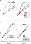
Growth of parental strains with inactivating lesions in amtB and suppressor strains at low NH3. Strains were grown at pH 5.5 with 0.5 mm NH4Cl as the nitrogen source and 0.1% glucose as the carbon source at 37°. All strains that grew more slowly than wild type reached a slightly higher final OD420 for reasons that we have not explored. (A) Strains with C-terminal lesions were wild-type (NCM3722), AmtBΔC-term (NCM4199), amtB null (amtB∷Spc, NCM4310), AmtBL394A (NCM4238), and AmtBfs (NCM4275). (B) Strains with lesions in cytoplasmic loop residues were AmtBE121A (NCM4348), AmtBR122A (NCM4349), AmtBK184A (NCM4326), AmtBR185A (NCM4327), AmtBK190A (NCM4344), AmtBE191A (NCM4346), and AmtBK255A (NCM4350). AmtBE121A, Y404N (NCM4474) was a suppressor strain. (C) Suppressor strains with the shortest C-terminal truncations or with missense mutations were AmtBdouble, Y404stop (NCM4278), AmtBR185A, Y404stop (NCM4471), AmtBdouble, A403D (NCM4281), AmtBL394A, A403D (NCM4382), and AmtBfs, R384L (NCM4424). (D) A suppressor strain with an hflB lesion was AmtBL394A HflBG361S (NCM4387). The suppressor strain with an hflC lesion was NCM4405 (HflCV80M AmtBfs), and the strains with hflC and hflK deletions were NCM4777 (ΔhflC AmtBL394A) and NCM4776 (ΔhflK AmtBL394A), respectively. Control strains were NCM4770 (wild type) and NCM4774 (AmtBL394A). The data in A–D are from a single experiment that is representative of the experiments summarized in Tables 1, 3, and 4.
F<sc>igure</sc> 2.—
Figure 2.—
Uptake of [14C]MA by parental strains with inactivating lesions in amtB and suppressor strains. To induce amtB expression, strains were grown with glutamine as the sole nitrogen source at pH 7 and at 37°. (A) Strains were wild-type (NCM3722), AmtBΔC-term (NCM4199), AmtBL394A (NCM4238), AmtBfs (NCM4275), and amtB null (NCM4310). Assays were performed at 37°. (B) Strains were wild type (NCM3722) and HflBG361S, AmtBL394A (NCM4387). They were assayed at both 37° and room temperature. The data in A and B are from a single experiment that is representative of findings made in at least three independent trials.
F<sc>igure</sc> 3.—
Figure 3.—
Locations of inactivating C-terminal and loop lesions in AmtB and of suppressor lesions in the C terminus. (A) Sequence of E. coli AmtB. Transmembrane (TM) segments (Khademi et al. 2004) are in black boxes. The first TM segment of AmtB (TM0) has been removed to leave a total of 11 TM segments. TM1-5 and TM6-10 are pseudosymmetric and the long TM11 crosses the protein diagonally. The C-terminal tail and odd-numbered loops are in the cytoplasm. When GlnK was bound to AmtB and the C terminus was ordered, the beginning of TM6 was shortened by one helical turn and loop 5 was extended (Conroy et al. 2007). Inactivating lesions in the C-terminal tail or loop residues are in pink. Positions at which the C-terminal tail makes contacts with loop residues are indicated above the sequence. Contacts within a monomer are in orange and contacts with the adjacent monomer are in blue. At every 10th residue, there is a tick under the sequence. (B) Locations of suppressor lesions in the C terminus. Missense lesions are in green and truncations are indicated with carats above the sequence. Residues that make hydrogen bonds to loop residues of the same monomer or the adjacent monomer are indicated with an “X” below the sequence.
F<sc>igure</sc> 4.—
Figure 4.—
Locations of missense suppressors in the C-terminal tail of AmtB. A space-filling model of the cytoplasmic face of E. coli AmtB was created using PyMol from Protein Data Bank entry 2NUU deposited by Conroy et al. (2007). The C-terminal tail of the light gray monomer is in brown and green, and loop 5 of the same monomer is in pink. Positions at which missense suppressors of inactivating C-terminal lesions or loop lesions were obtained—R384, A403, and Y404—are in green, and loop residues at which alanine substitutions inactivated AmtB—E121, R122, R185, and K255—are in blue. Only E121 and K255 are clearly visible. The other two monomers are in medium gray with dark gray C-terminal tails.
F<sc>igure</sc> 5.—
Figure 5.—
Locations of suppressor lesions in hflB. Approximate positions of lesions are indicated by ticks below the diagram. The diagram is adapted from Figure 1 of Ito and Akiyama (2005). HflB is divided into three regions: an initial membrane-bound region of two transmembrane-spanning segments (TMs) separated by a large periplasmic loop (Peri); an ATPase domain (AAA+ ATPase), which includes the second region of homology characteristic of AAA ATPases (SRH); a protease domain, which contains an α-helical coiled region (Coil). The domains are separated by a narrow space. Some conserved sequence motifs are shown at the top with residues known to be functionally important in boldface type. The Walker A motif in the original figure has been corrected to match the text. Whereas E479 was proposed to be the third Zn2+ ligand (Ito and Akiyama 2005), a subsequent publication indicated that D495 was the third ligand and that HflB was a unique Asp_Zincin protease (Bieniossek et al. 2006).
F<sc>igure</sc> 6.—
Figure 6.—
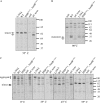
Effect of an hflB lesion on the behavior of AmtBL394A in vitro. Proteins were labeled to steady state with [35S]methionine, and AmtB was isolated with polyclonal antiserum raised against AmtB-his6 as described in materials and methods. Prior to electrophoresis, immunoprecipitated AmtB was incubated in LDS sample buffer containing 50 mm DTT for 1 hr at different temperatures—0°, 35°, 45°, 55°, and 90°, as indicated. Strains were wild type (NCM3722), AmtBΔC-term (NCM4199), AmtBL394A (NCM4238), HflBG361S AmtBL394A (NCM4387), and amtB null (NCM4590). (A) The gel was run for 2.5 rather than 1 hr to increase separation of full-length AmtB from AmtBΔC-term. Positions of the two trimers are indicated with arrows to the left of the gel. (B) Positions of monomers of full-length AmtB and AmtBΔC-term are indicated with arrows to the left of the gel. (C) Positions of AmtB aggregates are indicated with a bracket to the left of the gel. Positions of trimers of full-length AmtB and AmtBΔC-term (lower) are indicated with arrows. The gel shows that there is very little monomer at disaggregation temperatures up to 55°. Positions of Benchmark Prestained Protein Ladder standards (Invitrogen) are shown to the right of the gels in A–C, and dotted lines indicate positions at which the images of lanes were removed from the gels. Data shown are from a single experiment and are representative of findings made in three independent pulse-chase/immunoprecipitation trials.
F<sc>igure</sc> 7.—
Figure 7.—
View of the cytoplasmic face of AmtB and hypothetical diagram of the effects of amtB and hflB mutations. (Left) The space-filling model is the same as that in Figure 4. The cytoplasmic C-terminal tails are dark and the three monomers are in different shades of gray. The tails cross from one monomer to another (counterclockwise) to complete the cytoplasmic vestibule of the adjacent monomer (Conroy et al. 2007; Gruswitz et al. 2007). The large dots indicate centers of the pores. (Right) Approximate two-dimensional projection of the left image. The only side chain indicated is Y404. The red carat denotes the L394A mutation in the kink of the C-terminal tail. The HflB protease/chaperone “tacks” or “locks” the damaged tail to the adjacent monomer to inactivate AmtB. Apparently, it is positioned much like the wild-type tail (see text). In the absence of HflB, the tail is released from the adjacent monomer, as indicated by a dotted line, and behaves as if it were deleted, i.e., ≈ AmtBΔC-term. Arrows indicate single mutagenic events.

References

    1. Akiyama, Y., A. Kihara, H. Tokuda and K. Ito, 1996. FtsH (HflB) is an ATP-dependent protease selectively acting on SecY and some other membrane proteins. J. Biol. Chem. 271 31196–31201. - PubMed
    1. Andrade, S. L., and O. Einsle, 2007. The Amt/Mep/Rh family of ammonium transport proteins. Mol. Membr. Biol. 24 357–365. - PubMed
    1. Andrade, S. L., A. Dickmanns, R. Ficner and O. Einsle, 2005. Crystal structure of the archaeal ammonium transporter Amt-1 from Archaeoglobus fulgidus. Proc. Natl. Acad. Sci. USA 102 14994–14999. - PMC - PubMed
    1. Baba, T., T. Ara, M. Hasegawa, Y. Takai, Y. Okumura et al., 2006. Construction of Escherichia coli K-12 in-frame, single-gene knockout mutants: the Keio collection. Mol. Syst. Biol. 2 2006–0008. - PMC - PubMed
    1. Bieniossek, C., T. Schlach, M. Bumann, M. Meister, R. Meier et al., 2006. The molecular architecture of the metalloprotease FtsH. Proc. Natl. Acad. Sci. USA 103 3066–3071. - PMC - PubMed

Publication types

MeSH terms