. 2009 Aug;16(8):890-6.
doi: 10.1038/nsmb.1627. Epub 2009 Jul 13.

Diversity of chemical mechanisms in thioredoxin catalysis revealed by single-molecule force spectroscopy

Affiliations

Diversity of chemical mechanisms in thioredoxin catalysis revealed by single-molecule force spectroscopy

Raul Perez-Jimenez et al. Nat Struct Mol Biol. 2009 Aug.

Erratum in

  • Nat Struct Mol Biol. 2009 Dec;16(12):1331. Gaucher, Eric [corrected to Gaucher, Eric A]

Abstract

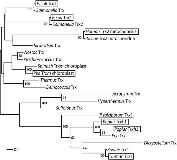
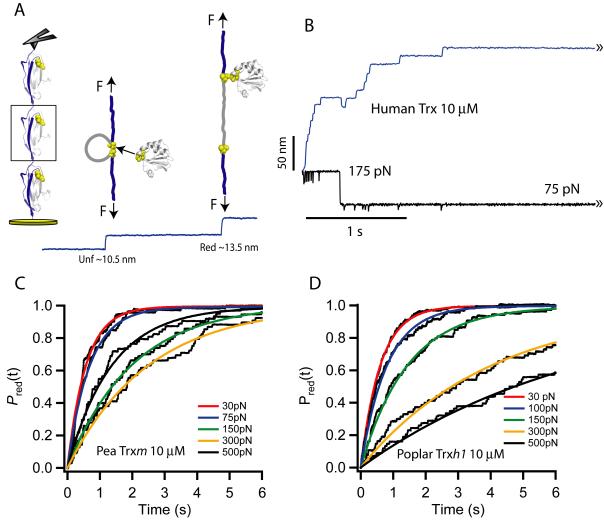
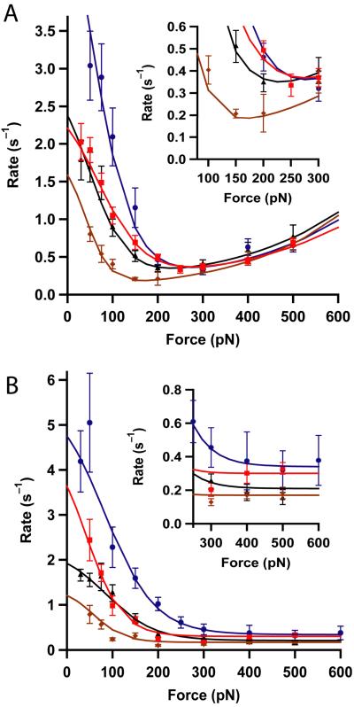
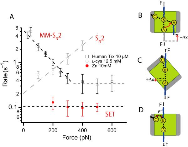
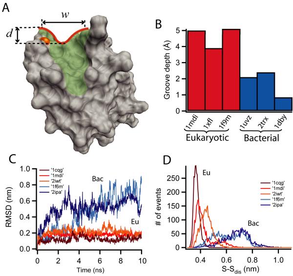
Thioredoxins (Trxs) are oxidoreductase enzymes, present in all organisms, that catalyze the reduction of disulfide bonds in proteins. By applying a calibrated force to a substrate disulfide, the chemical mechanisms of Trx catalysis can be examined in detail at the single-molecule level. Here we use single-molecule force-clamp spectroscopy to explore the chemical evolution of Trx catalysis by probing the chemistry of eight different Trx enzymes. All Trxs show a characteristic Michaelis-Menten mechanism that is detected when the disulfide bond is stretched at low forces, but at high forces, two different chemical behaviors distinguish bacterial-origin from eukaryotic-origin Trxs. Eukaryotic-origin Trxs reduce disulfide bonds through a single-electron transfer reaction (SET), whereas bacterial-origin Trxs show both nucleophilic substitution (S(N)2) and SET reactions. A computational analysis of Trx structures identifies the evolution of the binding groove as an important factor controlling the chemistry of Trx catalysis.


Figures

Figure 1
Figure 1. Phylogeny of Trx homologs from representative species of the three domains of life
Branch lengths were estimated using maximum likelihood with rate variation modeled according to a gamma distribution. Scale bar represents amino acid replacements per site per unit evolutionary time. Posterior probabilities are shown at nodes of the phylogeny when greater than 50%. The lack of strong node supports deep in the phylogeny results from the ambiguous placement of mitochondrial sequences, possibly due to long-branch attraction effects with non-bacterial sequences. In contrast, there is strong support for the grouping of chloroplast and cyanobacteria (not shown). Boxes highlight the proteins experimentally studied in this work.
Figure 2
Figure 2. Single molecule force-clamp detection of disulfide bond reduction events catalyzed by thioredoxin enzymes
(A) Graphic representation of the force-clamp experiment. A first force pulse rapidly unfolds the I27 modules of a polyprotein, exposing the buried disulfide bonds to the solvent. A second force pulse monitors single-disulfide reduction events which are uniquely identified by the extension of the residues trapped behind the disulfide bond. (B) Trace showing the unfolding and consequent disulfide reductions of a (I27G32C-A75C)8 polyprotein. In the example shown the unfolding pulse was set at 175 pN for 0.3 s and the reduction pulse was set to 75 pN for several seconds. (C) Summed and averaged traces of disulfide bond reductions at different forces (second pulse) for pea Trxm (10 μM) and (D) for poplar Trxh1 (10 μM). The smooth curves are single-exponential fits from which we measure the rate of reduction as r = 1/τ, where τ is the time constant measured by the fits at each given force.
Figure 3
Figure 3. Force-dependency of the rate of disulfide reduction by Trx enzymes from different species
(A) Bacterial-origin Trxs: human mitochondrial Trx2 (blue), E. coli Trx1 (red), pea chloroplastic Trxm (black), E. coli Trx2 (brown). While the Michaelis-Menten (low force) mechanism differs in magnitude among the Trxs, the simple SN2 like reaction observed at higher forces is very similar in all of them. The inset shows a magnified view of the traces in the region where they reach a minimum. (B) Eukaryotic-origin Trxs: human Trx1 (blue, from ref 11), Plasmodium Trx (red), poplar Trxh1 (black), and poplar Trxh3 (brown). The most noticeable feature is the absence of the SN2 like reaction at high forces in all eukaryotic Trxs. The inset shows an expansion of the minimum rate of reduction attained at high forces. In all experiments the concentration of Trx was 10 μM. The smooth lines are fits of the kinetic model described in the supplementary information. The kinetics parameters obtained are summarized in Table 1. The error bars are given by the standard error of the mean obtained with the bootstrap method.
Figure 4
Figure 4. The three chemical mechanisms of disulfide reduction detected by force-clamp spectroscopy
A) Force dependency of the rate of reduction of disulfide bonds by different reducing agents. Human Trx (black squares, from ref 11) shows the characteristic enzymatic mechanism of reduction, marked by a negative force dependency that reaches a force independent minimum. L-cysteine (gray circles, from ref 12) shows the characteristic SN2 like mechanism marked by an exponential increase in the rate of reduction with the applied force. Finally, metallic Zn (red circles) demonstrates a reduction mechanism that appears force independent. Bacterial thioredoxin enzymes make use of all three mechanisms. B) Schematic representation of the Michaelis-Menten reduction mechanism present in all Trx enzymes. The substrate disulfide bond is shown in the binding groove of the enzyme. Rotation of the substrate disulfide bond against the pulling force is required for the 180° alignment with the catalytic cysteine, and for reduction to occur. C) We speculate that the simple SN2-like mechanism observed at high forces results from aligning the substrate disulfide bond with the catalytic cysteine, without entering the binding groove. This conformation is favored by a shallow binding groove allowing for the simple SN2-type lengthening of the substrate disulfide bond at the transition state, which is the origin of the exponential dependency of the rate of reduction. D) Representation of the single-electron transfer (SET) mechanism ubiquitous to all Trxs. This mechanism can occur irrespective of the orientation of the disulfide bond and is more visible in eukaryotic Trxs at high forces.
Figure 5
Figure 5. Structural analysis and molecular dynamics simulations of the binding groove in thioredoxin enzymes
(A) Geometric dissection of the hydrophobic binding groove, shaded in green and outlined in red for human Trx1 (pdb: 3trx). The depth and width of the binding groove in the region surrounding the catalytic cysteine are indicated by arrows and the active cysteine is colored in orange. (B) Groove depth for three eukaryotic-origin Trxs (red): human Trx1 (pdb:1mdi); A. thaliana Trxh1 (pdb:1xfl) and spinach Trxf (pdb:1f9m), and three bacterial-origin Trxs (blue): human Trx2 (pdb:1uvz); E. coli Trx1 (pdb:2trx) and C. reinhardtii Trxm (pdb:1dby). It is clear that the eukaryotic binding grooves are deeper than their bacterial counterparts. (C) Molecular dynamics simulation of the substrate mobility within the binding groove for different eukaryotic (red) and bacterial (blue) Trxs. Three eukaryotic complexes were used: human Trx with the substrate REF-1 (pdb:1cqg); human Trx with NF-κB (pdb:1mdi) and barley Trxh2 with protein BASI (pdb:2iwt). The two bacterial complexes were: E. coli Trx1 with Trx reductase, (pdb:1f6m) and B. subtilis Trx bound to ArsC (pdb:2ipa). The large RMSD of the bacterial Trxs (blue) indicates a high substrate mobility which may facilitate collisions orientated so as favoring SN2 reactions. Eukaryotic Trxs (red) are highly restricted which may explain the different chemical behavior at high forces as compared to bacterial Trxs (Fig.3). (D) Diatomic distance distribution of the S-S bond in the Trx-substrate mixed intermediate, using the same structures as in (C). Again we infer higher mobility of the substrate in bacterial-origin Trxs as indicated by the broader distance distributions of the bacterial complexes (blue) as compared to those of eukaryotic Trxs (red).

References

    1. Kraut DA, Carroll KS, Herschlag D. Challenges in enzyme mechanism and energetics. Annu Rev Biochem. 2003;72:517–71. - PubMed
    1. Henzler-Wildman KA, et al. Intrinsic motions along an enzymatic reaction trajectory. Nature. 2007;450:838–44. - PubMed
    1. Dai S, Friemann R, Glauser DA, Bourquin F, Manieri W, Schurmann P, Eklund H. Structural snapshots along the reaction pathway of ferredoxinthioredoxin reductase. Nature. 2007;448:92–6. - PubMed
    1. Mori T, Vale RD, Tomishige M. How kinesin waits between steps. Nature. 2007;450:750–4. - PubMed
    1. Asbury CL, Fehr AN, Block SM. Kinesin moves by an asymmetric hand-over-hand mechanism. Science. 2003;302:2130–4. - PMC - PubMed

Publication types

MeSH terms