

Morning glory syndrome associated with posterior lenticonus
- PMID: 19597560
- PMCID: PMC2708595
- DOI: 10.2174/1874205X00903010045
Morning glory syndrome associated with posterior lenticonus
Abstract
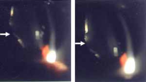
The clinical features of the morning glory syndrome (MSG) are demonstrated in a 12-year-old male patient with the posterior lenticonus in the left eye. This patient had retinal detachment in the left eye. A complete ocular examination was performed and the patient underwent a pars plana vitrectomy of the left eye. Slit-lamp examination revealed the posterior lenticonus with the posterior subcapsular opacities in the left eye. The fundus showed the symptoms of MGS. The discs were pink and deeply excavated, surrounded by a ring of chorioretinal pigmentary disturbance. The retina has remained reattached for six months after surgery. Although most cases of MGS present with retinal and vitrea abnormalities, it may also occur in association with the lens anomalies, including the posterior lenticonus and subcapsular cataract. This association may be helpful to explore the pathogenesis of MGS.
Keywords: Morning glory syndrome; posterior lenticonus..
Figures

References
-
- Kindler P. Morning glory syndrome: unusual congenital optic disk anomaly. Am J Ophthalmol. 1970;69(3):376–84. - PubMed
-
- Leitch RJ, Winter RM. Midline craniofacial defects and morning glory disc anomaly: a distinct clinical entity. Acta Ophthalmol Scand Suppl. 1996;219:16–9. - PubMed
-
- Gibbs ML, Jacobs M, Wilkie AO, Taylor D. Posterior lenticonus: clinical patterns and genetics. J Pediatr Ophthalmol Strabismus. 1993;30(3):171–5. - PubMed
-
- Colville DJ, Savige J. Alport syndrome: a review of the ocular manifestations. Ophthalmic Genet. 1997;18(4):161–73. - PubMed
-
- Jacobs K, Meire FM. Lenticonus. Bull Soc Belge Ophtalmol. 2000;277:65–70. - PubMed
LinkOut - more resources
Full Text Sources
Miscellaneous