. 2009;53(7):1097-104.
doi: 10.1387/ijdb.072489vb.

CBP/p300 and associated transcriptional co-activators exhibit distinct expression patterns during murine craniofacial and neural tube development

Affiliations

CBP/p300 and associated transcriptional co-activators exhibit distinct expression patterns during murine craniofacial and neural tube development

Vasker Bhattacherjee et al. Int J Dev Biol. 2009.

Abstract

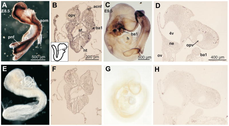
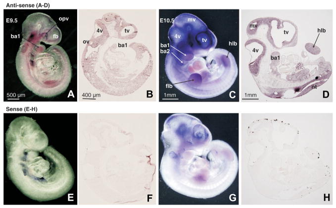
Mutations in each of the transcriptional co-activator genes - CBP, p300, Cited2, Cart1 and Carm1 - result in neural tube defects in mice. The present study thus furnishes a complete and comparative temporal and spatial expression map of CBP/p300 and associated transcriptional co-activators, Cited2, Cart1 and Carm1 during the period of murine neural tube development (embryonic days 8.5 to 10.5). Each co-activator except Cart1 was expressed in the dorsal neural folds on E8.5. Although CBP and p300 are functionally interchangeable in vitro, their respective expression patterns diverge during embryogenesis before neural fold fusion is complete. CBP gene expression was lost from the neural folds by E8.75 and was thereafter weakly expressed in the maxillary region and limb buds, while p300 exhibited strong expression in the first branchial arch, limb bud and telencephalic regions on E9.5. Cart1 exhibited strong expression in the forebrain mesenchyme from E9.0 through E10.5. Although CBP, p300, Carm1 and Cited2 share temporal expression on E8.5, these co-activators have different spatial expression in mesenchyme and/or the neuroepithelium. Nevertheless, co-localization to the dorsal neural folds on E8.5 suggests a functional role in elevation and/or fusion of the neural folds. Target genes, and pathways that promote cranial neural tube fusion that are activated by CBP/p300/Carm1/Cited2/Cart1-containing transcriptional complexes await elucidation.


Figures

Fig. 1
Fig. 1. Distribution of CREB binding protein (CBP) transcripts in E8.5 and E9.0 mouse embryos and tissue sections as determined by in situ hybridization
Mouse embryos (A,C,E,G) or tissue sections (B,D,F,H) were hybridized with CBP RNA antisense (A–D) or sense (E–H) probes. Panels showing E8.5 whole embryos are adjacent to stage-matched transverse sections (A–B, E–F), while E9.0 embryos are adjacent to stage-matched sagittal tissue sections of the of the anterior cranial region (C–D, G–H). The boxed inset in (B) shows a schematic representing the plane of section indicated by the dotted line. The boxed region of the E9.0 whole embryo (C) is shown in a sagittal tissue section (D). anf, anterior cranial neural folds: ba1/ba2, first/second branchial arch; cnf, cranial neural folds; fb, forebrain; h, heart; mv, mesencephalic vesicle; ne, neuroepithelium; nt, neural tube; opv, optic vesicle; plv, presumptive left ventricle; pnf, posterior neural folds.
Fig. 2
Fig. 2. Distribution of CREB binding protein (CBP) transcripts in E9.5 and E10.5 mouse embryos and tissue sections as determined by in situ hybridization
Mouse embryos (A,C,E,G) or tissue sections (B,D,F,H) were hybridized with CBP RNA antisense (A–D) or sense (E–H) probes. Panels showing E9.5 (A–B, E–F) and E10.5 (C–D, G–H) whole embryos are adjacent to panels showing the anterior regions of stage-matched sagittal sections. 4v, fourth ventricle; ba1/ba2, first/second branchial arch; flb, forelimb bud; h, heart; mv, mesencephalic vesicle; ne, neuroepithelium; opv, optic vesicle; tv, telencephalic vesicle.
Fig. 3
Fig. 3. Distribution of p300 protein expression in E8.5 and E9.5 mouse embryos as determined by whole-mount immunohistochemistry
E8.5 and E9.5 whole embryos (A,C) or sections (B,D) were incubated with anti-p300 polyclonal antibodies. Specificity of the antibody was demonstrated by preincubating the antibody with synthetic peptide before exposure to the embryos for one hour at 4°C at a 10 times molar excess (E–H). Note the exceptionally strong p300 signal emanating from the neural folds on E8.5. E8.5 embryos were not cleared (A,E), while E9.5 embryos were cleared (C,G) in order to optimally demonstrate p300 staining. Note that clearing causes the appearance of double images, such as the branchial arches on E9.5 (C) due to transparency of embryonic tissue. The boxed inset in (B) shows a schematic representing the plane of section as indicated by the dotted line. 4v, fourth ventricle; acnf, anterior cranial neural folds; af, anterior foregut; ba1, first branchial arch; h, heart; ne, neuroepithelium; nt, neural tube; opv, optic vesicle; ov, otic vesicle; pnf, posterior neural folds; som, somite.
Fig. 4
Fig. 4. Distribution of Cited2 transcripts in E9.5 and E10.5 mouse embryos and tissue sections as determined by in situ hybridization
E9.5 and E10.5 mouse embryos and sagittal sections were hybridized with the Cited2 RNA antisense probe (A–D) or with the corresponding sense probe as a negative control (E–H). 4v, fourth ventricle; ba1/2, first/second branchial arch; fb, forebrain; flb, forelimb bud; hlb, hindlimb bud; mv, mesencephalic vesicle; nt, neural tube; opv, optic vesicle; ov, otic vesicle; tv, telencephalic vesicle.
Fig. 5
Fig. 5. Distribution of Cart1 transcripts in E9.5 mouse embryos and tissue sections as determined by in situ hybridization
Mouse embryos or sections from E9.5 were hybridized with the Cart1 RNA antisense probe (A,B) or with the sense probe as a negative control (C,D). The expression detected in the orofacial region of the E9.5 embryo boxed in A was confirmed in the equivalent region of a sagittal section (B). ba1, first branchial arch; fb, forebrain; ne, neuroepithelium; opv, optic vesicle; tv, telencephalic vesicle.
Fig. 6
Fig. 6. Distribution of Cart1 transcripts in E10.5 mouse embryos and tissue sections as determined by in situ hybridization
E10.5 mouse embryos (A,E) or sections (B–D, F–H) were hybridized with the Cart1 RNA antisense probe (A–D) or with the sense probe as a negative control (EH). Panels (B–D, F–H) show adjacent sagittal sections of the cranial region from medial to lateral positions in the same embryo. 4v, fourth ventricle; ba1, first branchial arch; mnp, medial nasal process; mv, mesencephalic vesicle; opv, optic vesicle; tv, telencephalic vesicle.
Fig. 7
Fig. 7. Distribution of Carm1 transcripts in early mouse embryos at E8.5 and 9.5 as determined by in situ hybridization
Mouse embryos (A,C) or tissue sections (B,D) at E8.5- and E9.5 were hybridized with the Carm1 RNA antisense probe (A–D) or with the sense probe (E–H). The boxed inset in (B) shows a schematic representing the plane of section as indicated by the dotted line. (D) A cranial sagittal section of the E9.5 embryo. 4v, fourth ventricle; acnf, anterior cranial neural folds; ba1, first branchial arch; flb, forelimb bud; fn, frontonasal mesenchyme; nt, neural tube; ov, otic vesicle; opv, optic vesicle; plv, presumptive left ventricle; pnt, posterior neural tube.
Fig. 8
Fig. 8. Distribution of Carm1 transcripts in E10.5 mouse embryos and tissue sections as determined by in situ hybridization
Mouse embryos (A,C) or sagittal sections (B,D) at E10.5 were hybridized with the Carm1 RNA antisense probe (A,B) or with the sense probe (C,D). 4v, fourth ventricle; ba1, first branchial arch; fb, forebrain; flb, forelimb bud; hlb, hindlimb bud; tv, telencephalic vesicle.

References

    1. BAMFORTH SD, BRAGANCA J, ELORANTA JJ, MURDOCH JN, MARQUES FI, KRANC KR, FARZA H, HENDERSON DJ, HURST HC, BHATTACHARYA S. Cardiac malformations, adrenal agenesis, neural crest defects and exencephaly in mice lacking Cited2, a new Tfap2 co-activator. Nat Genet. 2001;29:469–474. - PubMed
    1. BARBERA JP, RODRIGUEZ TA, GREENE ND, WENINGER WJ, SIMEONE A, COPP AJ, BEDDINGTON RS, DUNWOODIE S. Folic acid prevents exencephaly in Cited2 deficient mice. Hum Mol Genet. 2002;11:283–293. - PubMed
    1. BRAGANCA J, ELORANTA JJ, BAMFORTH SD, IBBITT JC, HURST HC, BHATTACHARYA S. Physical and functional interactions among AP-2 transcription factors, p300/CREB-binding protein, and CITED2. J Biol Chem. 2003;278:16021–16029. - PubMed
    1. CHAKRAVARTI D, LAMORTE VJ, NELSON MC, NAKAJIMA T, SCHULMAN IG, JUGUILON H, MONTMINY M, EVANS RM. Role of CBP/P300 in nuclear receptor signalling. Nature. 1996;383:99–103. - PubMed
    1. CHEN SL, LOFFLER KA, CHEN D, STALLCUP MR, MUSCAT GE. The coactivator-associated arginine methyltransferase is necessary for muscle differentiation: CARM1 coactivates myocyte enhancer factor-2. J Biol Chem. 2002;277:4324–4333. - PubMed

Publication types

MeSH terms