. 2009 Nov 15;87(15):3428-37.
doi: 10.1002/jnr.22164.

Inactivation of fibroblast growth factor receptor signaling in myelinating glial cells results in significant loss of adult spiral ganglion neurons accompanied by age-related hearing impairment

Affiliations

Inactivation of fibroblast growth factor receptor signaling in myelinating glial cells results in significant loss of adult spiral ganglion neurons accompanied by age-related hearing impairment

S J Wang et al. J Neurosci Res. .

Abstract

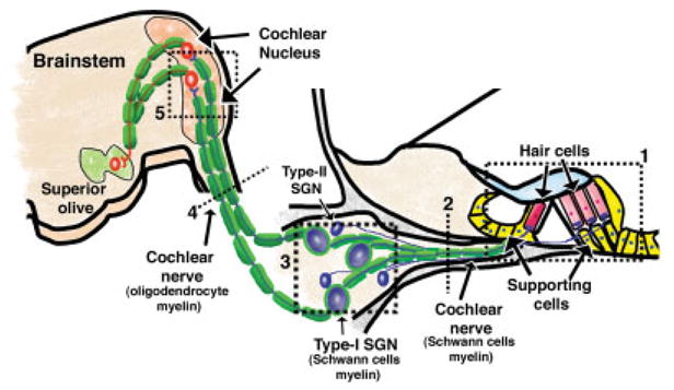
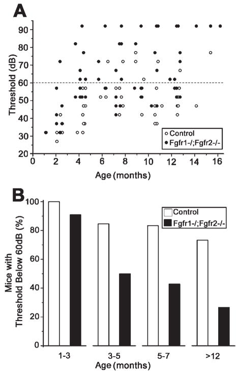
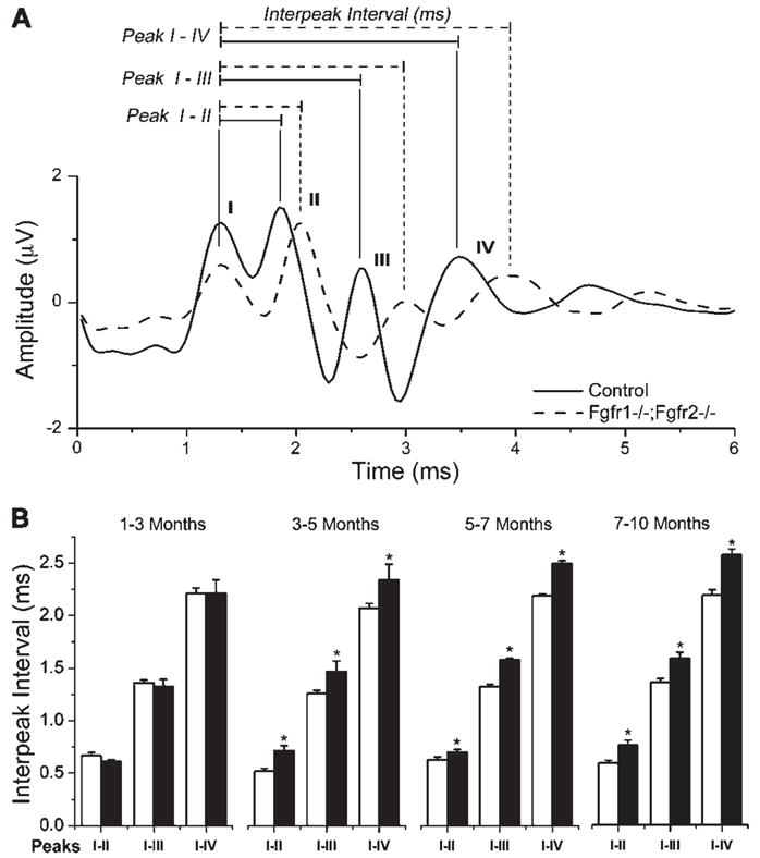
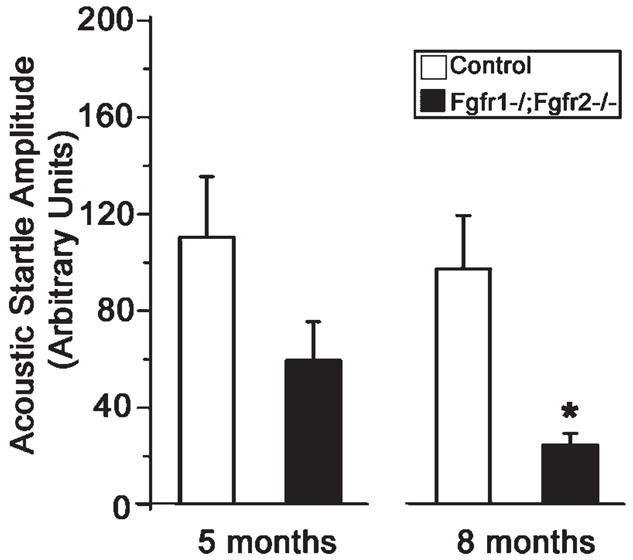
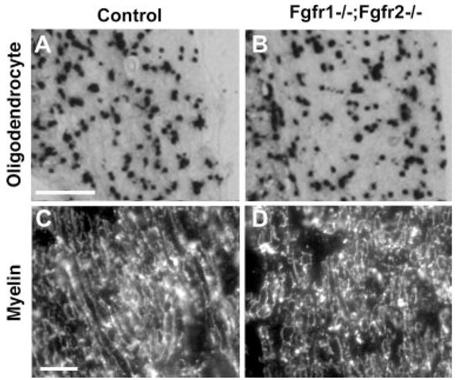
Hearing loss has been attributed to many factors, including degeneration of sensory neurons in the auditory pathway and demyelination along the cochlear nerve. Fibroblast growth factors (FGFs), which signal through four receptors (Fgfrs), are produced by auditory neurons and play a key role in embryonic development of the cochlea and in neuroprotection against sound-induced injury. However, the role of FGF signaling in the maintenance of normal auditory function in adult and aging mice remains to be elucidated. Furthermore, the contribution of glial cells, which myelinate the cochlear nerves, is poorly understood. To address these questions, we generated transgenic mice in which Fgfr1 and Fgfr2 were specifically inactivated in Schwann cells and oligodendrocytes but not in neurons. Adult mutant mice exhibited late onset of hearing impairment, which progressed markedly with age. The hearing impairment was accompanied by significant loss of myelinated spiral ganglion neurons. The pathology extended into the cochlear nucleus, without apparent loss of myelin or of the deletion-bearing glial cells themselves. This suggests that perturbation of FGF receptor-mediated glial function leads to the attenuation of glial support of neurons, leading to their loss and impairment of auditory functions. Thus, FGF/FGF receptor signaling provides a potentially novel mechanism of maintaining reciprocal interactions between neurons and glia in adult and aging animals. Dysfunction of glial cells and FGF receptor signaling may therefore be implicated in neurodegenerative hearing loss associated with normal aging.


Figures

Fig. 1
Fig. 1
CNP-Cre is selectively expressed in Schwann cells and oligodendrocytes but not neurons of the cochlear nucleus and spiral ganglion. Cross-sections of the peripheral segment of the cochlear nerve (A), spiral ganglion (B), and cochlear nucleus (C,D) from 2-week-old CNP-Cre-YFP-Rosa reporter mice were examined for the expression of yellow fluorescent protein (YFP). YFP was expressed by Schwann cells surrounding the cochlear nerve axons (*; A) and the SGNs (N; B) but was not detected in the axons (*) or neurons (N) themselves. Double labeling with APC (oligodendrocyte marker) and YFP showed complete overlap in the cochlear nucleus (C). In contrast, double labeling with NeuN (neuron marker) and YFP showed no overlap (D). Three mice were analyzed with similar results. Representative image is shown. Site at which sections were cut as referenced in Figure 2 are A: 2; B: 3; C,D: 5. Scale bars = 10 μm in A; 20 μm in B, insets; 100 μm in C,D.
Fig. 2
Fig. 2
Diagramatic representation of the regions of the auditory pathway investigated in the Fgfr1−/−; Fgfr2−/− mice. Within the cochlea is the organ of Corti (1), which contains hair cells and nonneuronal support cells. Inner and outer hair cells are synaptically connected through the myelinated and unmyelinated nerve fibers of the peripheral cochlear nerve, respectively (2), to the bipolar-spiral ganglion neurons (SGNs) in the spiral ganglion (3). Approximately 90% of SGNs are type I and myelinated by Schwann cells, and 10% are type II, which are unmyelinated. From the spiral ganglion, the cochlear nerve continues (4) to the CNS via the cochlear nucleus (5) in the brainstem and is myelinated by oligodendrocytes. The numbers show the sites at which the section was cut. These numbers are referenced in each figure. [Color figure can be viewed in the online issue, which is available at www.interscience.wiley.com.]
Fig. 3
Fig. 3
Fgfr1−/−; Fgfr2−/− mice exhibit progressive hearing loss with age. A: Adult mice were examined for hearing loss by auditory brainstem response (ABR) tests. Hearing sensitivity of individual control and mutant mice is shown as the thresholds of responses to a sound stimulus played at different intensities [in peak equivalent sound pressure level (peSPL), in dB], plotted as a function of age. Overall, mutant mice (solid circles) exhibited higher hearing thresholds than control mice (open circles) at all ages beyond 4 months. B: Mice that showed ABR response to stimuli below 60 dB peSPL were plotted as a percentage of total mice tested in each group. Animals with thresholds below 60 dB were considered to be within the boundary of “normal” hearing. Note the late onset of hearing impairment and progression with age in the mutant mice. Ten to fifteen mice were analyzed in each age group and genotype. Because no obvious differences were observed between males and females, data were pooled for the analysis.
Fig. 4
Fig. 4
Fgfr1−/−; Fgfr2−/− mice exhibit prolonged latency between successive peaks of ABR waveforms compared with controls. A: Representative ABR waveform showing amplitude in microvolts plotted against time in milliseconds. Four ABR peaks are shown from control (solid line) and mutant (dashed line) mouse recordings. In general, a delay in the interpeak interval was observed in mutants compared with controls for all peaks. In addition, reduced peak height is also observed. B: Four control (open bars) and four mutant mice (solid bars) were analyzed at approximately 2-month interval for IPI latencies over a period of 10 months. The average interpeak intervals from 87 dB, 82 dB, and 77 dB stimuli are plotted for peaks I–II, I–III, and I–IV in each age group. Note that mutants and controls displayed similar IPIs at 1–3 months of age, but mutants showed a significant delay (*P < 0.05) at older ages. Error bars represent SEM; N = 4.
Fig. 5
Fig. 5
Fgfr1−/−; Fgfr2−/− mice show a lower acoustic startle response than the control mice. Mice at approximately 5 and 8 months of age were examined for hearing loss by acoustic startle reflex measurements. Average startle amplitude from each of the 24 trials in which the startle stimulus was presented alone was calculated. Mean value of startle amplitude from both age group of mutant and control mice is shown. ANOVA revealed a significant effect of genotype, F(1,30) = 26.3, *P < .001. There was no interaction between these factors. A Tukey test revealed that mutant mice showed a smaller acoustic startle response than the control mice at both ages. Error bars represent SEM; N = 3–9.
Fig. 6
Fig. 6
Fgfr1−/−; Fgfr2−/− mice exhibit significant loss of type I spiral ganglion neurons and reactive gliosis in the cochlear nucleus. A: Nissl-stained sections of medial (a,b) and basal (c,d) turns of the cochlea, showing SGNs of 7-month-old control and Fgfr1−/−;Fgfr2−/− mice. A substantial loss of type I SGNs (large size, arrowhead) was observed, but not of type II SGNs (small size, arrow). Insets show magnified images of the cells denoted by arrows. B: Quantification of the numbers of type I SGNs in control (open bars) and mutant (solid bars) mice. Controls were set at baseline of 100%. Note the significant reduction (*P < 0.05) in the numbers of SGNs in the mutant apical, medial, and basal turns. Error bars represent SEM; N = 3 for all except apical and basal for controls, where N = 2. C: Transverse sections of the ventral cochlear nucleus from control (a) and mutant (b) mice were immunolabeled for glial fibrillary acidic protein (GFAP). Intense GFAP staining of thick astrocytic processes (arrowhead) and cell bodies (arrow), indicative of pathological gliosis, was observed in the mutants compared with controls. A representative experiment out of three is shown. Site at which sections were cut as referenced in Figure 2, A: 3; C: 5. Scale bars = 100 μm in A; 50 μm in inset; 20 μm in C.
Fig. 7
Fig. 7
Peripheral glial cells and peripheral myelin appear similar in Fgfr1−/−; Fgfr2−/− and control mice. Sections of cochlea, immunolabeled with Myo7a (a marker of hair cells), showed that inner and outer hair cells (IHC and OHC) were present in the organ of Corti in both control and Fgfr1−/−; Fgfr2−/− mice (A,B). Similarly, phase-contrast microscopy showed that support cells were also present in the control and mutants (C,D, solid arrowheads). Expression of MBP (a marker for myelin and myelinating Schwann cells) showed a similar pattern of immunolabeling of the spiral ganglion (SG) and peripheral segment of cochlear nerve in mutant and control mice (E,F), further confirmed in cross-sections of cochlear nerves showing axons surrounded by MBP+ myelinating Schwann cells (insets in e,f). Cross-sections of spiral ganglions (G,H) immunostained with MBP show that spiral ganglion Schwann cells (SGSCs), ensheathing the spiral ganglion neurons (N) with myelin, are present in both the control and the Fgfr1−/−; Fgfr2−/− mice. Similar results were obtained when three or four mice of each genotype, ranging between 4 and 10 months of age were analyzed. Representative images are shown. Site at which sections were cut as referenced in Figure 2, A–D: 1; E–H: 3. Scale bars = 20 μm in a (applies to A,B); 20 μm in e (applies to E,F); 20 μm in in g (applies to g,h); 50 μm in c (applies to D,D); 5 μm in insets.
Fig. 8
Fig. 8
Oligodendrocytes and central myelin appear to be similar in Fgfr1−/−; Fgfr2−/− and control mice. Transverse sections of the ventral cochlear nucleus (A,B) or central segment of cochlear nerve (C,D) from control and mutant mice were analyzed by in situ hybridization for the oligodendrocyte marker PLP mRNA (A,B) or by immunohistochemistry for myelin marker MBP (C,D). Expression patterns of PLP mRNA and MBP were similar in control and Fgfr1−/−; Fgfr2−/− mice. Analysis of three or four mice of each genotype, ranging between 4 and 8 months of age, gave similar results. Representative images are shown. Site at which sections were cut as referenced in Figure 2, A,B: 5; C,D: 4. Scale bars = 200 μm in a (applies to A,B); 10 μm in c (applies to C,D).

Similar articles

Cited by

References

    1. Adams JC, Schulte BA. Histopathologic observations of the aging gerbil cochlea. Hear Res. 1997;104:101–111. - PubMed
    1. Armstrong RC, Le TQ, Frost EE, Borke RC, Vana AC. Absence of fibroblast growth factor 2 promotes oligodendroglial repopulation of demyelinated white matter. J Neurosci. 2002;22:8574–8585. - PMC - PubMed
    1. Bandtlow CE, Heumann R, Schwab ME, Thoenen H. Cellular localization of nerve growth factor synthesis by in situ hybridization. EMBO J. 1987;6:891–899. - PMC - PubMed
    1. Bansal R. Fibroblast growth factors and their receptors in oligodendrocyte development: implications for demyelination and remyelination. Dev Neurosci. 2002;24:35–46. - PubMed
    1. Bansal R, Kumar M, Murray K, Morrison RS, Pfeiffer SE. Regulation of FGF receptors in the oligodendrocyte lineage. Mol Cell Neurosci. 1996;7:263–275. - PubMed

Publication types

MeSH terms

Substances