

Insulin-like growth factor binding protein-7 induces activation and transdifferentiation of hepatic stellate cells in vitro
- PMID: 19598300
- PMCID: PMC2710780
- DOI: 10.3748/wjg.15.3246
Insulin-like growth factor binding protein-7 induces activation and transdifferentiation of hepatic stellate cells in vitro
Abstract
Aim: To investigate the role of insulin-like growth factor binding protein-7 (IGFBP-7) in the activation and transdifferentiation of hepatic stellate cells (HSC) in vitro.
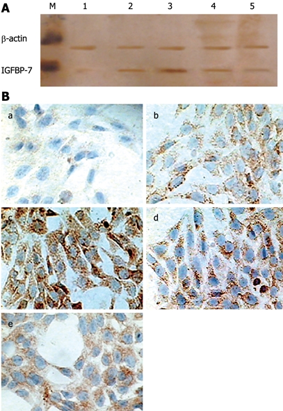
Methods: Rat HSC-T6 cells were cultured in separate dishes and treated with various concentration of transforming growth factor (TGF)-beta(1), IGFBP-7 or anti-IGFBP-7 antibody for 24 h. The supernatant or a cytoplasm suspension was obtained from cultured HSC, followed by transfer of cells to form cell-coated dishes. Immunocytochemistry and Western blotting were used to analyze the expression of IGFBP-7 induced by TGF-beta(1) and the level of fibronectin, collagen I and alpha-smooth muscle actin (SMA). The pro-apoptotic effect of anti-IGFBP-7 antibody was determined by flow cytometry.
Results: Immunocytochemistry and Western blotting revealed that the expression of IGFBP-7 in TGF-beta(1) treated HSC was significantly up-regulated compared to that in the control group. In addition, fibronectin, collagen I and alpha-SMA also showed enhanced expression in accordance with the transdifferentiation process in a dose-dependent manner to some extent. Moreover, flow cytometry suggested that anti-IGFBP-7 antibody induced apoptosis of activated HSC, which is responsible for the development of liver fibrosis, and may represent a novel pathway and target for therapeutic intervention.
Conclusion: IGFBP-7 showed increased expression in activated HSC and played an important role in the activation and transdifferentiation process of HSC. Anti-IGFBP-7 antibody may ameliorate liver fibrogenesis.
Figures


Similar articles
-
IGFBPrP1 induces liver fibrosis by inducing hepatic stellate cell activation and hepatocyte apoptosis via Smad2/3 signaling.World J Gastroenterol. 2014 Jun 7;20(21):6523-33. doi: 10.3748/wjg.v20.i21.6523. World J Gastroenterol. 2014. PMID: 24914373 Free PMC article.
-
Fluvastatin attenuates hepatic steatosis-induced fibrogenesis in rats through inhibiting paracrine effect of hepatocyte on hepatic stellate cells.BMC Gastroenterol. 2015 Feb 15;15:22. doi: 10.1186/s12876-015-0248-8. BMC Gastroenterol. 2015. PMID: 25886887 Free PMC article.
-
Transdifferentiation-dependent expression of alpha-SMA in hepatic stellate cells does not involve TGF-beta pathways leading to coinduction of collagen type I and thrombospondin-2.Matrix Biol. 2005 May;24(3):198-207. doi: 10.1016/j.matbio.2005.03.003. Matrix Biol. 2005. PMID: 15905080
-
Wnt signaling in liver fibrosis: progress, challenges and potential directions.Biochimie. 2013 Dec;95(12):2326-35. doi: 10.1016/j.biochi.2013.09.003. Epub 2013 Sep 13. Biochimie. 2013. PMID: 24036368 Review.
-
Cooperation of liver cells in health and disease.Adv Anat Embryol Cell Biol. 2001;161:III-XIII, 1-151. doi: 10.1007/978-3-642-56553-3. Adv Anat Embryol Cell Biol. 2001. PMID: 11729749 Review.
Cited by
-
Retrospective identification of intrinsic factors that mark pluripotency potential in rare somatic cells.bioRxiv [Preprint]. 2023 Feb 10:2023.02.10.527870. doi: 10.1101/2023.02.10.527870. bioRxiv. 2023. Update in: Cell Syst. 2024 Feb 21;15(2):109-133.e10. doi: 10.1016/j.cels.2024.01.001. PMID: 36798299 Free PMC article. Updated. Preprint.
-
Relationship of IGF-1 and IGF-Binding Proteins to Disease Severity and Glycemia in Nonalcoholic Fatty Liver Disease.J Clin Endocrinol Metab. 2021 Jan 23;106(2):e520-e533. doi: 10.1210/clinem/dgaa792. J Clin Endocrinol Metab. 2021. PMID: 33125080 Free PMC article. Clinical Trial.
-
Insulin-like Growth Factor Binding Proteins and Cellular Senescence Are Involved in the Progression of Non-Alcoholic Fatty Liver Disease and Fibrosis in a Mouse Model.Medicina (Kaunas). 2024 Mar 2;60(3):429. doi: 10.3390/medicina60030429. Medicina (Kaunas). 2024. PMID: 38541155 Free PMC article.
-
Differentiation of Adipose-Derived Stem Cells into Vascular Smooth Muscle Cells for Tissue Engineering Applications.Biomedicines. 2021 Jul 9;9(7):797. doi: 10.3390/biomedicines9070797. Biomedicines. 2021. PMID: 34356861 Free PMC article.
-
Role of IGFBP7 in Diabetic Nephropathy: TGF-β1 Induces IGFBP7 via Smad2/4 in Human Renal Proximal Tubular Epithelial Cells.PLoS One. 2016 Mar 14;11(3):e0150897. doi: 10.1371/journal.pone.0150897. eCollection 2016. PLoS One. 2016. PMID: 26974954 Free PMC article. Clinical Trial.
References
-
- Friedman SL. Liver fibrosis -- from bench to bedside. J Hepatol. 2003;38 Suppl 1:S38–S53. - PubMed
-
- Gressner AM. Liver fibrosis: perspectives in pathobiochemical research and clinical outlook. Eur J Clin Chem Clin Biochem. 1991;29:293–311. - PubMed
-
- Friedman SL. Molecular regulation of hepatic fibrosis, an integrated cellular response to tissue injury. J Biol Chem. 2000;275:2247–2250. - PubMed
-
- Parsons CJ, Takashima M, Rippe RA. Molecular mechanisms of hepatic fibrogenesis. J Gastroenterol Hepatol. 2007;22 Suppl 1:S79–S84. - PubMed
Publication types
MeSH terms
Substances
LinkOut - more resources
Full Text Sources
Other Literature Sources
Miscellaneous
