. 2009 Jul 14:4:27.
doi: 10.1186/1749-8104-4-27.

Chick Lrrn2, a novel downstream effector of Hoxb1 and Shh, functions in the selective targeting of rhombomere 4 motor neurons

Affiliations

Chick Lrrn2, a novel downstream effector of Hoxb1 and Shh, functions in the selective targeting of rhombomere 4 motor neurons

Laura C Andreae et al. Neural Dev. .

Abstract

Background: Capricious is a Drosophila adhesion molecule that regulates specific targeting of a subset of motor neurons to their muscle target. We set out to identify whether one of its vertebrate homologues, Lrrn2, might play an analogous role in the chick.

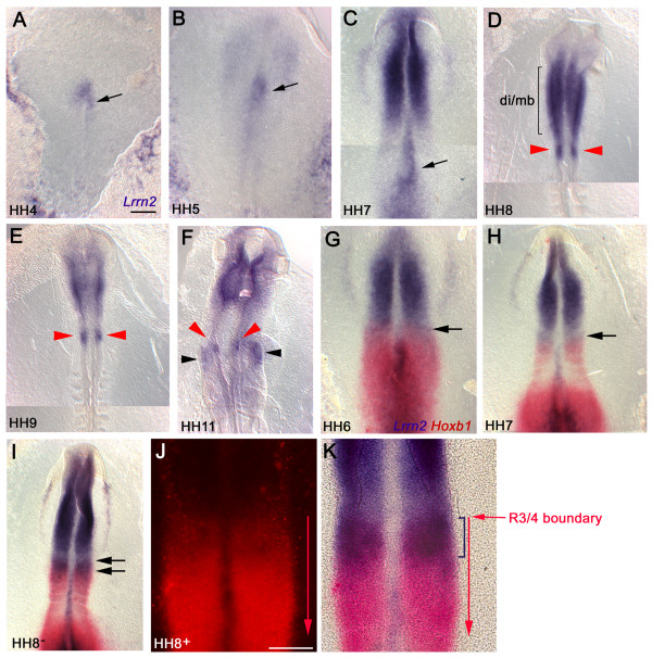
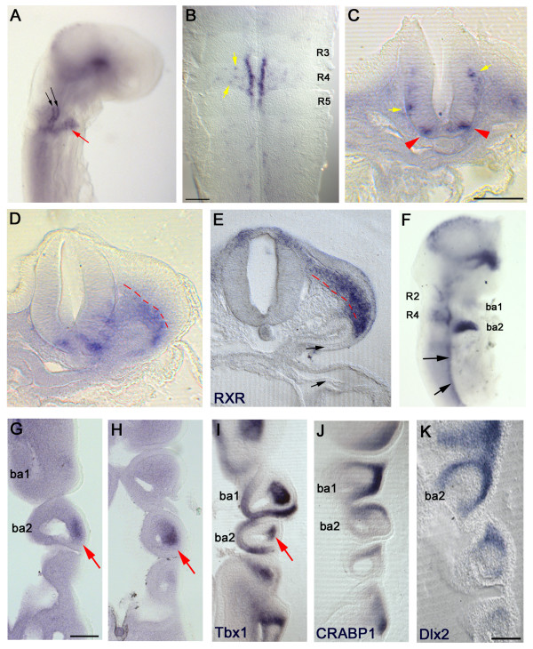
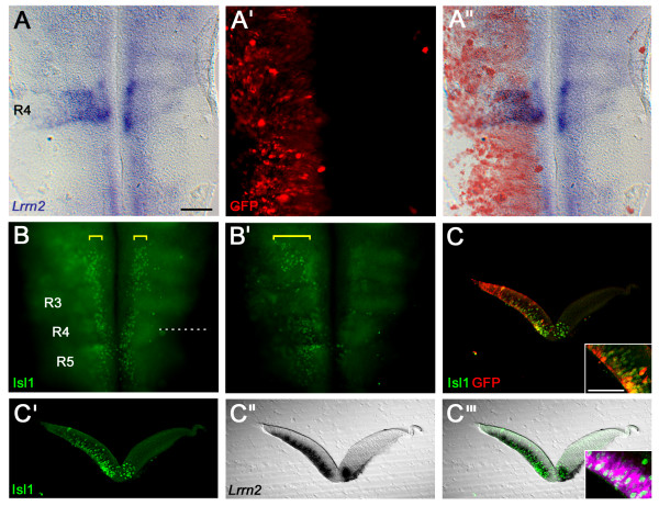
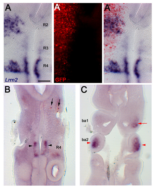
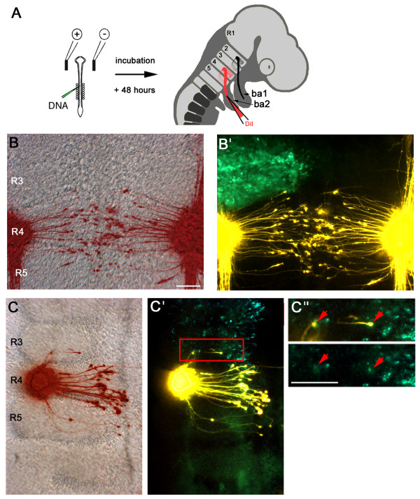
Results: We have shown that Lrrn2 is expressed from early development in the prospective rhombomere 4 (r4) of the chick hindbrain. Subsequently, its expression in the hindbrain becomes restricted to a specific group of motor neurons, the branchiomotor neurons of r4, and their pre-muscle target, the second branchial arch (BA2), along with other sites outside the hindbrain. Misexpression of the signalling molecule Sonic hedgehog (Shh) via in ovo electroporation results in upregulation of Lrrn2 exclusively in r4, while the combined expression of Hoxb1 and Shh is sufficient to induce ectopic Lrrn2 in r1/2. Misexpression of Lrrn2 in r2/3 results in axonal rerouting from the r2 exit point to the r4 exit point and BA2, suggesting a direct role in motor axon guidance.

Conclusion: Lrrn2 acts downstream of Hoxb1 and plays a role in the selective targeting of r4 motor neurons to BA2.


Figures

Figure 1
Figure 1
Time-course of Lrrn2 expression. Chick embryos from HH4–11 following in situ hybridisation for Lrrn2 and Hoxb1. All views are dorsal side up, anterior to the top. (A) HH4, (B) HH5, (C) HH7. Note asymmetric expression of Lrrn2 on the right side of the node (arrows in (A-C)). (D) HH8, (E) HH 9, (F) HH11. Expression of Lrrn2 starts to localise to the r4 region at HH8 and by HH9 is clearly distinguishable (red arrowheads in (D, E)). Strong staining is also seen in the prospective diencephalon-midbrain region (di/mb) (D). At HH11, r4 expression within the neuroepithelium can be seen (red arrowheads), along with expression in the adjacent mesoderm (black arrowheads in (F)). (G-K) Double in situ hybridisation with Lrrn2 (blue) and Hoxb1 (red). (G) HH6, (H) HH7; arrows indicate anterior boundary of Hoxb1 expression (future r3/4 boundary). (I) HH8-: the region between the double, black arrows highlights overlap in expression, corresponding to presumptive r4. (J) HH8+: high power view of hindbrain region with fluorescent image of Hoxb1 expression where the r3/4 boundary is clear, despite some quenching due to the NBT/BCIP staining of Lrrn2, and (K) brightfield image showing the same. The anterior boundary of the Lrrn2 stripe (bracket) coincides with the anterior boundary of Hoxb1, indicating that Lrrn2 is expressed in early r4. Scale bars: 100 μm.
Figure 2
Figure 2
Lrrn2 marks rhombomere 4 motor neurons and the second branchial arch. In situ hybridisation with (A-D, F-H) Lrrn2, (E) RXR, (I) Tbx1, (J) CRABP1, (K) Dlx2 at HH14 (A-E) and HH18 (F-K). (A) Wholemount embryo shows Lrrn2 expression in two stripes in ventral r4 (black arrows) and in nearby cells within the presumptive BA2 region (red arrow). (B) Flatmounted hindbrain shows Lrrn2 expression restricted to two columns immediately alongside the ventral midline of r4, with a few scattered cells within the body of r4 (yellow arrows). (C) Transverse section through the hindbrain at the level of r4 shows expression in the outer layer (red arrowheads). Yellow arrows indicate more dorsal cells seen also in (B). (D) Next posterior transverse section of the same series as (C) shows staining in the ventrally located mesoderm (below red dashed line). (E) Neural crest marker RXR labels a dorsally migrating population lying above the same line. (F) At HH18, Lrrn2 is strongly expressed in BA2 but is essentially absent from BA1. Staining is also visible in scattered cells more dorsally in r2 and r4, as well as in the ventral motor column (black arrows) and ventral midbrain. (G, H) Adjacent coronal sections through the branchial arch region at HH18 shows Lrrn2 in the central core area of BA2 (red arrows). (I) Tbx1 expression labels the mesodermal core of the branchial arches; staining in the BA2 core area is indicated by a red arrow. (J) CRABP1 and (K) Dlx2 are neural crest markers and show a peripheral crescent of staining in the branchial arches, unlike Lrrn2. Scale bars: 100 μm (E, I-K). Courtesy of Robyn Quinlan and Anthony Graham.
Figure 3
Figure 3
Shh upregulates Lrrn2 in rhombomere 4. (A-A") Co-electroporation of Shh and GFP results in overexpression throughout the left side of the hindbrain as visualised by anti-GFP labelling, reflecting a similar pattern of Shh misexpression (data not shown). This results in an upregulation of Lrrn2 expression only in r4. (A) Lrrn2 expression, (A') GFP expression, (A") overlay. (B, B') The same embryo, following immunocytochemistry with anti-Isl1/2 antibody (green) to label motor neurons, showing the normal pattern of Isl1/2 antibody staining in motor neurons lying adjacent to the floor plate (yellow brackets); (B) photo taken with focal plane set ventrally, and (B') a more dorsal focal plane showing upregulation of Isl1/2 throughout the left side of the hindbrain, as one would predict (expanded yellow bracket). Dashed line indicates the plane of section of (C-C"'). (C-C"') Transverse sections through the hindbrain at the level of r4. (C) Overlay of Isl1/2 in green and GFP in red, indicating unilateral Shh overexpression and induction of Isl1/2 expression. Inset shows high power image. (C'-C"') The expansion of Lrrn2 expression can be seen to be confined to the mantle layer (C") and colocalises with upregulation of Islet1/2 (Isl1/2 alone (C'); overlay with Lrrn2 (C"'); inset shows high power view with Lrrn2 expression pseudocoloured in magenta to more easily visualise overlap with Isl1/2 staining). Scale bar: 100 μm; 50 μm (inset).
Figure 4
Figure 4
Hoxb1 regulates Lrrn2 expression. (A-A") Overlay to show co-expression of ectopic Shh and Hoxb1 and their effect on Lrrn2 expression in a flatmounted hindbrain, using an anti-GFP antibody to localise the region of misexpression. Widespread overexpression is seen throughout the hindbrain, but Lrrn2 is upregulated in r1, r2 and r4 only. (A) Brightfield view of Lrrn2 expression and (A') fluorescence of anti-GFP antibody. (B-C) Coronal sections through an embryo electroporated with RCASBP(B)-mHoxb1 at HH10 and processed 2 days later for Lrrn2 (dark purple) and mouse Hoxb1 (red) expression by in situ hybridisation. (B) Section taken through ventral r4 show endogenous expression of Lrrn2 in r4 (black arrowheads) and ectopic expression of mouse Hoxb1 (black arrows). (C) Corresponding section taken more ventrally through the branchial arches of the same embryo shows upregulation of Lrrn2 expression in BA1 mesoderm (red arrow) and endogenous expression in BA2 (red arrowheads). Scale bar: 100 μm.
Figure 5
Figure 5
Ectopic Lrrn2 can cause misrouting of axons. (A) Schematic to show experimental approach. On the left is shown a HH10 chick embryo with electrodes positioned on either side of the hindbrain. DNA is injected into the neural tube and following electroporation is expressed unilaterally on the side of the positive electrode. After 48 h embryos are harvested and the retrograde dye DiI is injected into BA2. Dye travels along axonal processes and labels the cell bodies from which they originate. (B, B') Flatmounted hindbrain from a control embryo electroporated with pCAβ-eGFPm5 shows motor projections to BA2 labelled by retrograde DiI injection. (B) Overlay of artificially coloured DiI fluorescence (red) showing BA2 axons and a brightfield photograph of the hindbrain. (B') Overlay showing strong GFP expression in r2 and r3 on the left side of the hindbrain. All axons projecting to BA2 clearly originate in r4 and r5 with no cell bodies lying anterior to the r3/4 boundary. (C, C') Embryo with misexpression of Lrrn2-GFP in r2/3 shows several ectopic axons that project from cell bodies in r3 to BA2. (C") High power views of the boxed region in (C') showing that the ectopic axons originate from Lrrn2-GFP+ cells (red arrows). Scale bars: 100 μm.

References

    1. Lumsden A, Krumlauf R. Patterning the vertebrate neuraxis. Science. 1996;274:1109–1115. doi: 10.1126/science.274.5290.1109. - DOI - PubMed
    1. Hunt P, Gulisano M, Cook M, Sham MH, Faiella A, Wilkinson D, Boncinelli E, Krumlauf R. A distinct Hox code for the branchial region of the vertebrate head. Nature. 1991;353:861–864. doi: 10.1038/353861a0. - DOI - PubMed
    1. Murphy P, Davidson DR, Hill RE. Segment-specific expression of a homoeobox-containing gene in the mouse hindbrain. Nature. 1989;341:156–159. doi: 10.1038/341156a0. - DOI - PubMed
    1. Wilkinson DG, Bhatt S, Cook M, Boncinelli E, Krumlauf R. Segmental expression of Hox-2 homoeobox-containing genes in the developing mouse hindbrain. Nature. 1989;341:405–409. doi: 10.1038/341405a0. - DOI - PubMed
    1. Alexandre D, Clarke JD, Oxtoby E, Yan YL, Jowett T, Holder N. Ectopic expression of Hoxa-1 in the zebrafish alters the fate of the mandibular arch neural crest and phenocopies a retinoic acid-induced phenotype. Development. 1996;122:735–746. - PubMed

Publication types

MeSH terms

Substances

LinkOut - more resources