. 2009 Jul 14:4:28.
doi: 10.1186/1749-8104-4-28.

Mathematical modeling supports substantial mouse neural progenitor cell death

Affiliations

Mathematical modeling supports substantial mouse neural progenitor cell death

Michael J McConnell et al. Neural Dev. .

Abstract

Background: Existing quantitative models of mouse cerebral cortical development are not fully constrained by experimental data.

Results: Here, we use simple difference equations to model neural progenitor cell fate decisions, incorporating intermediate progenitor cells and initially low rates of neural progenitor cell death. Also, we conduct a sensitivity analysis to investigate possible uncertainty in the fraction of cells that divide, differentiate, and die at each cell cycle.

Conclusion: We demonstrate that uniformly low-level neural progenitor cell death, as concluded in previous models, is incompatible with normal mouse cortical development. Levels of neural progenitor cell death up to and exceeding 50% are compatible with normal cortical development and may operate to prevent forebrain overgrowth as observed following cell death attenuation, as occurs in caspase 3-null mutant mice.


Figures

Figure 1
Figure 1
Appropriate ventricular zone (VZ) output requires greater than 5% neural progenitor cell (NPC) death. (A, B) Two simple cell decision paradigms are used to identify a plausibility window and calculate viable levels of NPC death. Beginning at 'P' and following the arrows clockwise, in model DG1 (A) the NPC population Pi-1 is doubled at the ith mitosis, then NPC death, 'D,' and differentiation, 'Q,' are imposed, so that the P population for the subsequent cell cycle (CC) is given by Pi = 2(1 - di - qi)Pi-1. (B) In model DG2 dying NPCs are removed from the NPC population prior to doubling the population, and Pi = (1 - qi)2(1 - di)Pi-1. (C, D) Model output from P0 = 550,000. VZ output (total Q cells, y-axis) is plotted after 11 CCs when the indicated fraction of NPCs die at each CC (Death, x-axis). The window of plausible VZ output, between 1 × 10⁷ and 2.72 × 10⁷, is indicated by dashed lines. (C) Using model DG1, the center of the plausible VZ output range (total Q cells = 1.86 × 10⁷) corresponds to 19% NPC death, as indicated by the solid line that descends from plotted VZ output to the x-axis. (D) Similarly, the center of the plausibility window using model DG2 corresponds to 17% NPC death.
Figure 2
Figure 2
Model DG1 is insensitive to perturbations in qi and di. (A) Most (>90%) model realizations give plausible ventricular zone (VZ) output (gray bars). The distribution of 10,000 simulations of VZ output (where formula image = 0.19 for all i) is plotted as a histogram. Simulation outputs follow a log-normal distribution. The solid vertical line indicates the geometric population mean, and dashed vertical lines indicate 1 and 2 standard deviations from the mean. (B) Three examples of points in 22-dimensional parameter space where plausible VZ output is observed. The q fractions are plotted along a dashed line and d fractions are plotted along a solid line. The upper panel is from a bin below the mean, the center panel is from the bin at the mean, and the lower panel is from a bin above the mean. CC, cell cycle.
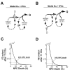
Figure 3
Figure 3
Intermediate progenitor cells (IPCs) modestly increase the requirement for neural progenitor cell (NPC) death. (A, B) IPCs (IP) are incorporated into (A) model DG1 and (B) model DG2 similarly. A fraction of Q cells (q') become IPCs, undergo one CC (2 *IP), then re-enter the Q population. (C, D) Ventricular zone (VZ) output plotted as described in Figure 1. (C) In model DG1 the plausibility window is centered at 22% NPC death. (D) In model DG2 the plausibility window is centered at 24% NPC death.
Figure 4
Figure 4
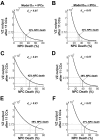
Expansion cell cycles permit much more neural progenitor cell (NPC) death at later cell cycles (CCs). Ventricular zone (VZ) output plotted as described in Figure 1. During expansion CCs, di = 0.05; NPC death during subsequent CCs is indicated on the x-axis. (A, B) After three expansion CCs the plausibility window is centered at 29% using model DG1 (A) and 24% using model DG2 (B). (C, D) After four expansion CCs the plausibility window is centered at 37% using model DG1 (C) and 28% using model DG2 (D). (E, F) After five expansion CCs the plausibility window is centered at 53% using model DG1 (E) and 36% using model DG2 (F).
Figure 5
Figure 5
Constrained models require substantial levels of progenitor cell (NPC) death. Ventricular zone (VZ) output as plotted in Figure 1, including intermediate progenitor cells (IPCs) as in model DG1 (Figure 3A) and model DG2 (Figure 3B). (A, B) After three expansion cell cycles (CCs) the plausibility window is centered at 33% using model DG1 (A) and 33% using model DG2 (B). (C, D) After four expansion CCs the plausibility window is centered at 42% using model DG1 (C) and 39% using model DG2 (D). (E, F) After five expansion CCs the plausibility window is centered at 59% using model DG1 (E) and 47% using model DG2 (F).

References

    1. Gotz M, Huttner WB. The cell biology of neurogenesis. Nat Rev Mol Cell Biol. 2005;6:777–788. doi: 10.1038/nrm1739. - DOI - PubMed
    1. Capela A, Temple S. LeX is expressed by principle progenitor cells in the embryonic nervous system, is secreted into their environment and binds Wnt-1. Dev Biol. 2006;291:300–313. doi: 10.1016/j.ydbio.2005.12.030. - DOI - PubMed
    1. Fishell G, Kriegstein A. Cortical development: new concepts. Neuron. 2005;46:361–362. doi: 10.1016/j.neuron.2005.04.016. - DOI - PubMed
    1. Bystron I, Blakemore C, Rakic P. Development of the human cerebral cortex: Boulder Committee revisited. Nat Rev Neurosci. 2008;9:110–122. doi: 10.1038/nrn2252. - DOI - PubMed
    1. Angevine JB, Sidman RL. Autoradiographic study of cell migration during histogenesis of the cerebral cortex of the mouse. Nature. 1961;192:766–768. doi: 10.1038/192766b0. - DOI - PubMed

Publication types