. 2009 Jul 14:2:44.
doi: 10.1186/1755-8794-2-44.

Novel transcriptional profile in wrist muscles from cerebral palsy patients

Affiliations

Novel transcriptional profile in wrist muscles from cerebral palsy patients

Lucas R Smith et al. BMC Med Genomics. .

Abstract

Background: Cerebral palsy (CP) is an upper motor neuron disease that results in a progressive movement disorder. Secondary to the neurological insult, muscles from CP patients often become spastic. Spastic muscle is characterized by an increased resistance to stretch, but often develops the further complication of contracture which represents a prominent disability in children with CP. This study's purpose is to characterize alterations of spastic muscle on the transcriptional level. Increased knowledge of spastic muscle may lead to novel therapies to improve the quality of life for children with CP.

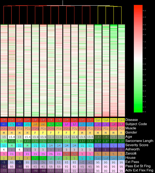
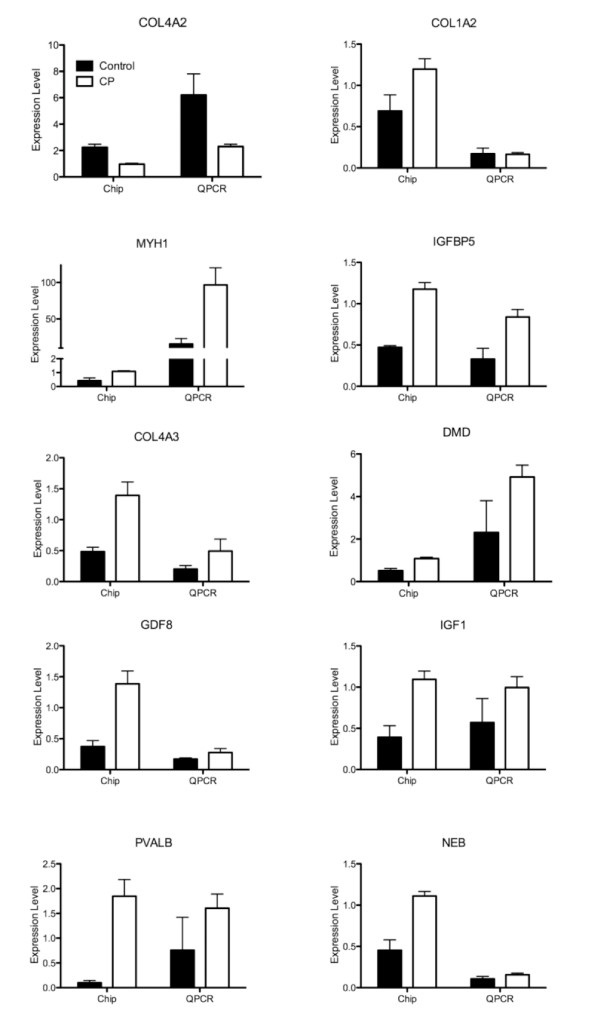
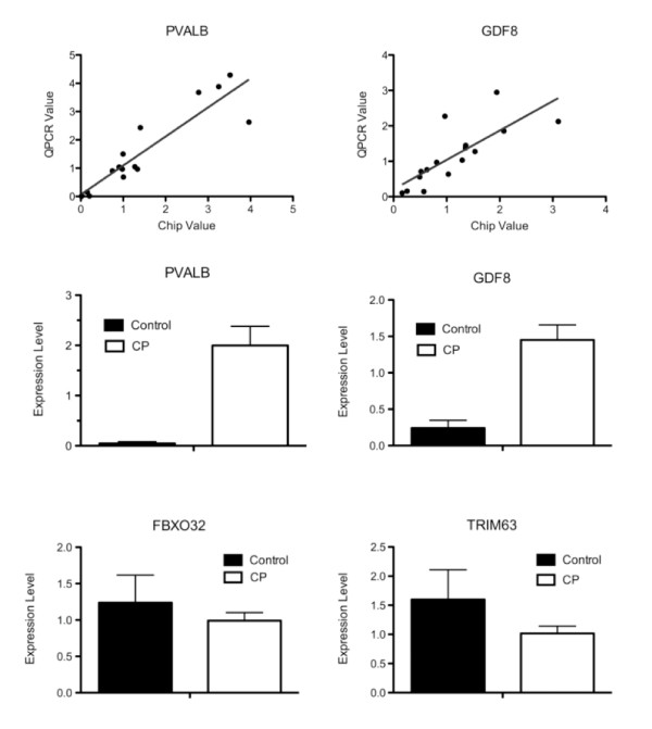
Method: The transcriptional profile of spastic muscles were defined in children with cerebral palsy and compared to control patients using Affymetrix U133A chips. Expression data were verified using quantitative-PCR (QPCR) and validated with SDS-PAGE for select genes. Significant genes were determined using a 2 x 2 ANOVA and results required congruence between 3 preprocessing algorithms.

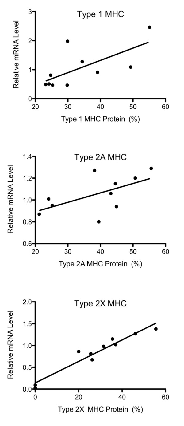
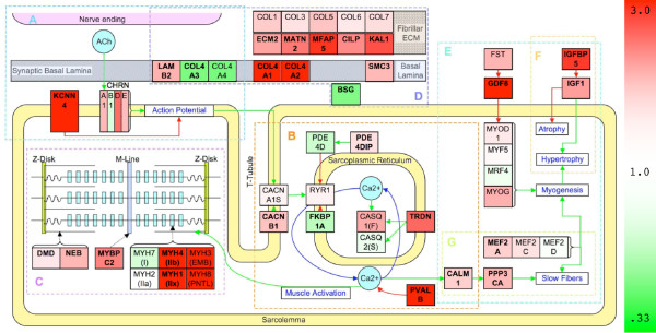
Results: CP patients clustered independently and 205 genes were significantly altered, covering a range of cellular processes. Placing gene expression in the context of physiological pathways, the results demonstrated that spastic muscle in CP adapts transcriptionally by altering extracellular matrix, fiber type, and myogenic potential. Extracellular matrix adaptations occur primarily in the basal lamina although there is increase in fibrillar collagen components. Fiber type is predominately fast compared to normal muscle as evidenced by contractile gene isoforms and decrease in oxidative metabolic gene transcription, despite a paradoxical increased transcription of slow fiber pathway genes. We also found competing pathways of fiber hypertrophy with an increase in the anabolic IGF1 gene in parallel with a paradoxical increase in myostatin, a gene responsible for stopping muscle growth. We found evidence that excitation-contraction coupling genes are altered in muscles from patients with CP and may be a significant component of disease.

Conclusion: This is the first transcriptional profile performed on spastic muscle of CP patients and these adaptations were not characteristic of those observed in other disease states such as Duchenne muscular dystrophy and immobilization-induced muscle atrophy. Further research is required to understand the mechanism of muscle adaptation to this upper motor neuron lesion that could lead to the development of innovative therapies.


Figures

Figure 1
Figure 1
Top: Condition tree created using Pearson Correlation for the similarity score and an average linkage clustering algorithm. The tree was created based on all present features. MAS5 data were used with expression values normalized to each features median. Features are ordered from highest expression ratio to lowest. Bottom: Clinical conditions color-coded with values for each sample.
Figure 2
Figure 2
QPCR results compared to GeneChip results for several individual genes shown from CP (open bars) and CTRL (filled bars) patients. Error bars represent SEM. QPCR data are from dilute (1:100) samples to test multiple genes and represent transcript level relative to total RNA (fg/μg). GeneChip data are normalized to the median value for each gene and averaged across CP or CTRL samples.
Figure 3
Figure 3
(A/B) Sample-to-sample correlation between the QPCR and GeneChip results. (A) PVALB, parvalbumin; (B) GDF8, myostatin. The solid line is a best fit regression line. (C-F) QPCR results showing the difference between CP and CTRL patients for specific genes, (C) PVALB, (D) GDF8, (E) FBXO32/MaFBX/Atrogin, (F) TRIM63/MuRF. Values are expressed determined relative to GAPDH and normalized to the median value for each individual gene. (*) represents significant difference (P < 0.05). Error bars represent SEM.
Figure 4
Figure 4
Comparison of the GeneChip mRNA data to protein SDS-PAGE gel data for three myosin heavy chains commonly expressed in human skeletal muscle. A: Type 1 MHC (MYH6), B: Type 2A MHC (MYH2), C: Type 2X MHC (MYH1). mRNA data are normalized to the median value for each gene on the chip using MAS5 preprocessing and protein data are normalized to total MyHC content.
Figure 5
Figure 5
Pathways specific to muscle analysis of transcription in CP muscle. Pathways A-G involved in muscle function. Color is determined by the expression ratio. Up-regulated genes are red and down-regulated genes are green. Green connectors represent activation and red connectors represent inhibition in the direction of the arrow. Bolded genes represent those that are significantly altered in all three preprocessing algorithms. Italic genes (RAS, MAPK) are sets of genes involved in muscle MAPK pathway in muscle, but are not colored by expression because many individual genes are applicable and not altered in CP. Pathways represented are A: NMJ, B: ECC, C: MC, D: ECM, E: MYG, F: FT, and G: IGF1.

References

    1. Rosenbaum P, Paneth N, Leviton A, Goldstein M, Bax M, Damiano D, Dan B, Jacobsson B. A report: the definition and classification of cerebral palsy April 2006. Dev Med Child Neurol Suppl. 2007;109:8–14. - PubMed
    1. Johnson A. Cerebral palsies: epidemiology and causal pathways. Archives of Disease in Childhood. 2000;83:279A. doi: 10.1136/adc.83.3.279a. - DOI - PMC - PubMed
    1. Hoon AH., Jr Neuroimaging in cerebral palsy: Patterns of brain dysgenesis and injury. Journal of child neurology. 2005;20:936–939. doi: 10.1177/08830738050200120201. - DOI - PubMed
    1. Korzeniewski SJ, Birbeck G, DeLano MC, Potchen MJ, Paneth N. A systematic review of neuroimaging for cerebral palsy. Journal of child neurology. 2008;23:216–227. doi: 10.1177/0883073807307983. - DOI - PubMed
    1. Wood E. The child with cerebral palsy: diagnosis and beyond. Seminars in pediatric neurology. 2006;13:286–296. doi: 10.1016/j.spen.2006.09.009. - DOI - PubMed