. 2009 Aug 1;69(15):6331-8.
doi: 10.1158/0008-5472.CAN-08-4329. Epub 2009 Jul 14.

CCL5-mediated endogenous antitumor immunity elicited by adoptively transferred lymphocytes and dendritic cell depletion

Affiliations

CCL5-mediated endogenous antitumor immunity elicited by adoptively transferred lymphocytes and dendritic cell depletion

Yolanda Nesbeth et al. Cancer Res. .

Abstract

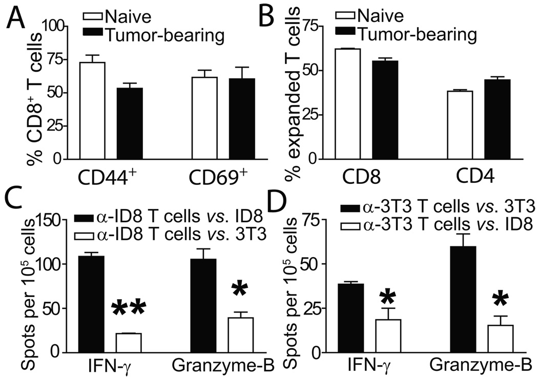
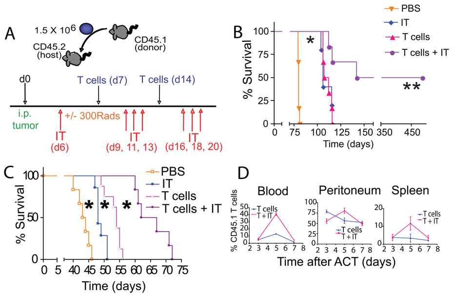
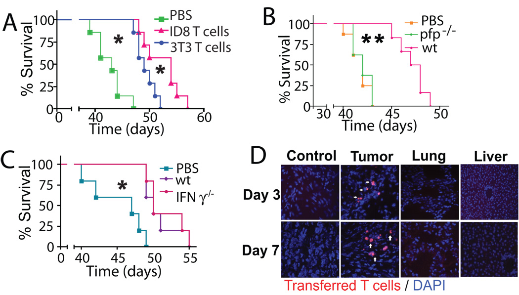
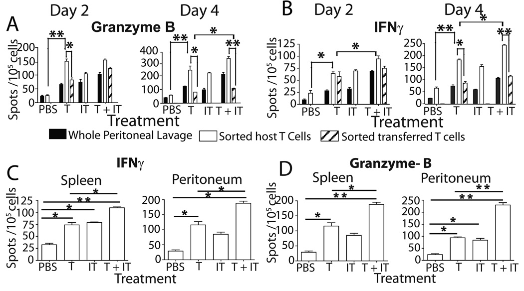
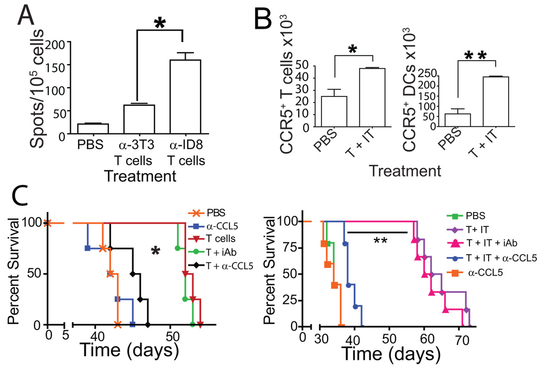
Adoptive transfer of antitumor T cells is a promisingly effective therapy for various cancers, but its effect on endogenous antitumor immune mechanisms remains largely unknown. Here, we show that the administration of naive T cells de novo primed for only 7 days against tumor antigens resulted in the durable rejection of otherwise lethal ovarian cancers when coupled with the depletion of tumor-associated immunosuppressive dendritic cells (DC). Therapeutic activity required tumor antigen specificity and perforin expression by the adoptively transferred T cells, but not IFN-gamma production. Importantly, these shortly primed T cells secreted large amounts of CCL5, which was required for their therapeutic benefit. Accordingly, transferred T cells recruited CCR5(+) DCs into the tumor, where they showed distinct immunostimulatory attributes. Activated CCR5(+) host T cells with antitumor activity also accumulated at tumor locations, and endogenous tumor-specific memory T cells remained elevated after the disappearance of transferred lymphocytes. Therefore, persistent, long-lived antitumor immunity was triggered by the administration of ex vivo activated T cells, but was directly mediated by immune cells of host origin. Our data unveil a CCL5-dependent mechanism of awakening endogenous antitumor immunity triggered by ex vivo expanded T cells, which is augmented by tumor-specific targeting of the cancer microenvironment.


Figures

Figure 1
Figure 1. Phenotypic characterization of tumor-reactive and irrelevantly primed T cells
(A) Activation status of T cells from either healthy (naive) or ID8-Defb29/Vegf-a tumor-bearing (tumor-bearing) mice primed to ID8-Defb29/Vegf-a antigens. Representative of 3 independent experiments. (B) T cells expanded from tumor-bearing or healthy mice show comparable proportions of CD8 vs. CD4 T cells. (C) IFN-γ and granzyme-B ELISPOT analyses of naïve T cells primed to ID8-Defb29/Vegf-a antigens, in response to their cognate tumor antigen (α-ID8 T cells vs. ID8), or NIH-3T3 fibroblasts (α-ID8 T cells vs. 3T3). Representative of 2 independent experiments (n=6/group, total). (D) Similar ELISPOT analysis performed with naïve T cells primed to irrelevant NIH-3T3 fibroblasts (*- P<0.05; **- P<0.01).
Figure 2
Figure 2. Elimination of CD11c+ cells enhances the efficacy of ACT
(*- P<0.05; **- P<0.01; T cells - T cell transfer; IT - anti-CD11c immunotoxin). (A) Experimental design. CD45.2+mice were inoculated i.p. with tumor and treated on day 7 and day 14 of tumor progression with 1.5×10⁶ CD45.1+ tumor-primed T cells (T cells). When relevant, tumor-bearing mice were irradiated on the first day of T cell transfer. Tumor-bearing mice were depleted of CD11c cells with the anti-CD11c immunotoxin (IT) the day prior to the initial T cell transfer and thrice weekly for two weeks thereafter. (B) ACT plus anti-CD11c immunotoxin, but not individual treatments, induced the regression of established intraperitoneal ID8-luciferase tumors (n=12 per group in 2 independent experiments). (C) ID8-Defb29/Vegf-a tumor-bearing mice receiving ACT plus IT survived significantly longer than untreated mice, or mice receiving individual treatments (n=24 mice/group, total in 4 independent experiments). (D) In vivo proliferation of transferred congenic (CD45.1+) T cells with (T+IT) or without (T) immunotoxin administration. The percentage of transferred cells at specific locations over time was determined (n=8 mice/group in 2 independent experiments).
Figure 3
Figure 3. The therapeutic effects of ACT require perforin and antigen specific priming but not IFNγ
(*- P<0.05; **- P<0.01). (A) Survival of ID8-Defb29/Vegf-a tumor–bearing mice receiving T cells primed either against ID8-Defb29/Vegf-a tumor cell antigens (“ID8 T cells”) or irrelevant NIH-3T3 fibroblasts (“3T3 T cells”) was established (n=8 mice/group). (B) ID8-Defb29/Vegf-a–tumor expanded splenic T cells from CD45.2+ perforin deficient (pfp−/−) or wild-type (wt) mice were transferred into ID8-Defb29/Vegf-a tumor-bearing, congenic (CD45.1) mice (n=16 mice/group in 2 independent experiments). Survival was determined. (C) Survival induced by IFN-γ deficient (IFN-γ−/−) T cells (n=16 mice/group in 2 independent experiments) was compared in an identical experiment. Mice were not irradiated in (B) or (C). (D) Mice bearing 14-day old flank ID8-Defb29/Vegf-a tumors received ID8-Defb29/Vegf-a primed red fluorescent T cells intraperitoneally and immunofluorescent microscopy was performed. (magnification - 200 X; n=6 mice/group).
Figure 4
Figure 4. Adoptive T cell therapy induces host immune responses that are augmented by the elimination of CD11c+ cells
(* - P<0.05; **- P<0.01; T- T cell transfer; IT- anti-CD11c immunotoxin). (A) Beginning on day 2 after T cell transfer, peritoneal host (CD45.2) and transferred (CD45.1) T cells were independently FACS sorted. Whole peritoneal wash samples, sorted host cells and sorted transferred cells (n=6/group in 2 independent experiments) were then analyzed by ELISPOT for secretion of granzyme-B (left) and IFN-γ (right) in response to tumor antigen. (B) IFN-γ (left) and granzyme-B (right) ELISPOT analyses were performed on peritoneal wash samples and spleens obtained from mice at day 26 of tumor progression, when transferred T cells are no longer detectable (n=9/group in 3 independent experiments).
Figure 5
Figure 5. Expanded T cells secrete CCL5 and activate host immune cells
(*- P<0.05; **-P<0.01, Mann-Whitney) T- T cell transfer; IT - anti-CD11c immunotoxin. (A) Supernatants from naïve splenic T cells primed for 7-days against ID8-Defb29/Vegf-a tumor cell antigens were examined by Luminex assays for cytokine production. (B) (upper) Phenotypic analysis of host (CD45.2+CD3+) T cells accumulated at the tumor site 3 days after ACT. (lower) Expression profile of host CD11c+ (DCs) at tumor sites 3 days after treatment. Values represent mean percentages +/− SEM. (right) Quantification of MHCII+CD80+CD11c+ DCs in the peritoneum of mice treated with ACT and anti-CD11c immunotoxin (T+IT), compared to control mice. (C) Luminex analysis of PMA/Ionomycin-stimulated peritoneal MHCII+CD80+CD11c+ DCs from pre and post-treatment mice. (D) Proliferation of CFSE-labeled magnetically purified (left) allogeneic Balb/c slenocytes (proliferation indices-2.84 (treated) vs 1.07 (untreated)) and (center) CD8+ OT-1 splenocytes (proliferation indices-4.68 (treated) vs 1.56 (untreated)) upon culture with sorted peritoneal MHCII+CD80+CD11c+ DCs from pre (shaded) and post-treatment (unshaded) mice. (right) Granzyme-B ELISPOT analysis of CD3+ splenocytes from untreated tumor-bearing mice cultured with sorted peritoneal MHCII+CD80+CD11c+ DCs from pre and post-treatment mice. n=9 in 3 independent experiments.
Figure 6
Figure 6. Tumor specific priming and CCL5 are required for successful ACT
(A) ELISPOT analysis of the number of sorted endogenous (CD45.2+) T cells producing IFN-γ in response to tumor antigen 7 days after ACT with tumor-specific or irrelevantly primed T cells (n=6 mice/group, total for A – B in 2 independent experiments). (B) Adoptive transfer of tumor-reactive T cells following anti-CD11c immunotoxin administration (T+IT) increases the total number of host CCR5+ (CD45.2+) CD3+ T cells and CD11c+ DCs (n=6 in 2 independent experiments). (C) Administration of a neutralizing antibody to CCL5 concurrently with ACT, but not an irrelevant IgG, diminished the ability of transferred T cells to increase survival of tumor bearing mice in the presence (right) or absence (left) of CD11c cell depletion. (P<0.05, n=12 in 2 independent experiments).

References

    1. Rosenberg SA, Restifo NP, Yang JC, Morgan RA, Dudley ME. Adoptive cell transfer: a clinical path to effective cancer immunotherapy. Nat Rev Cancer. 2008;8:299–308. - PMC - PubMed
    1. Dudley ME, Wunderlich JR, Robbins PF. Cancer regression and autoimmunity in patients after clonal repopulation with antitumor lymphocytes. Science. 2002;298:850–854. - PMC - PubMed
    1. Morgan RA, Dudley ME, Wunderlich JR. Cancer regression in patients after transfer of genetically engineered lymphocytes. Science. 2006;314:126–129. - PMC - PubMed
    1. Hunder NN, Wallen H, Cao J. Treatment of metastatic melanoma with autologous CD4+ T cells against NY-ESO-1. N Engl J Med. 2008;358:2698–2703. - PMC - PubMed
    1. Klebanoff CA, Gattinoni L, Restifo NP. CD8+ T-cell memory in tumor immunology and immunotherapy. Immunol Rev. 2006;211:214–224. - PMC - PubMed

Publication types