. 2009 Oct;8(10):2368-81.
doi: 10.1074/mcp.M900188-MCP200. Epub 2009 Jul 14.

In planta proteomics and proteogenomics of the biotrophic barley fungal pathogen Blumeria graminis f. sp. hordei

Affiliations

In planta proteomics and proteogenomics of the biotrophic barley fungal pathogen Blumeria graminis f. sp. hordei

Laurence V Bindschedler et al. Mol Cell Proteomics. 2009 Oct.

Abstract

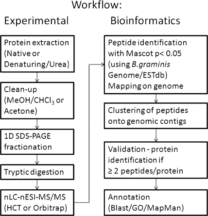
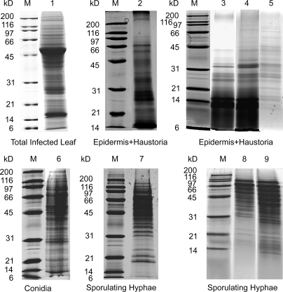
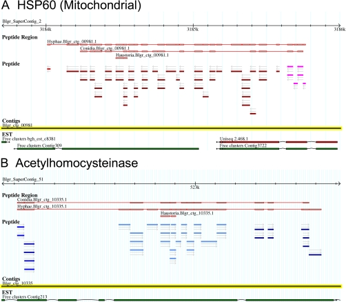
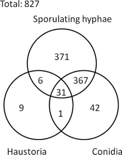
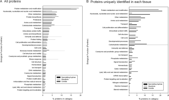
To further our understanding of powdery mildew biology during infection, we undertook a systematic shotgun proteomics analysis of the obligate biotroph Blumeria graminis f. sp. hordei at different stages of development in the host. Moreover we used a proteogenomics approach to feed information into the annotation of the newly sequenced genome. We analyzed and compared the proteomes from three stages of development representing different functions during the plant-dependent vegetative life cycle of this fungus. We identified 441 proteins in ungerminated spores, 775 proteins in epiphytic sporulating hyphae, and 47 proteins from haustoria inside barley leaf epidermal cells and used the data to aid annotation of the B. graminis f. sp. hordei genome. We also compared the differences in the protein complement of these key stages. Although confirming some of the previously reported findings and models derived from the analysis of transcriptome dynamics, our results also suggest that the intracellular haustoria are subject to stress possibly as a result of the plant defense strategy, including the production of reactive oxygen species. In addition, a number of small haustorial proteins with a predicted N-terminal signal peptide for secretion were identified in infected tissues: these represent candidate effector proteins that may play a role in controlling host metabolism and immunity.


Figures

Fig. 1.
Fig. 1.
Images of the three different B. graminis f. sp. hordei structures analyzed in this study. A, micrograph of mature ungerminated conidia. The conidia were isolated from sporulating colonies, suspended in water, and photographed using differential interference contrast microscopy (bar, 10 μm). B, scanning electron micrograph of fungal mycelium (sporulating hyphae) growing epiphytically on the barley leaf surface. The “runner hyphae” adhering tightly to the leaf surface are visible at the edge of the colony; the aerial conidiophores emerge from the center of the colony (bar, 100 μm). C, low magnification epifluorescence microscopy image of haustorial cells within epidermal cells of heavily infected primary leaves of barley. Epiphytic structures pictured in B were removed, and the leaf epidermis was dissected. The fungal haustoria (h) and the plant stomata (st) are labeled. The fungal structures were stained by Alexa Fluor 488 bound to wheat germ agglutinin and visualized using a FITC emission/barrier filter (bar, 20 μm). D, close-up micrograph of haustorial structures inside the plant cells. The sample is the same as in C (bar, 10 μm).
Fig. 2.
Fig. 2.
Diagram of the experimental and bioinformatics work flow summarizing the main processes used in the analysis of the samples as described in this study.
Fig. 3.
Fig. 3.
Images of barley/Blumeria protein extracts separated by SDS-PAGE and stained with colloidal Coomassie. Each gel lane was sliced into 2-mm bands for in-gel digestion with trypsin prior to analysis by mass spectrometry. Lane 1, soluble proteins (60 μg) extracted in native conditions from TL tissue of infected plants (10 dpi). Lanes 2–9, proteins extracted under denaturing conditions with a urea-containing buffer following clean-up and concentration by either TA or CM precipitation. Lanes 2–5, barley/Blumeria EH protein extracts prepared from infected barley leaves (10 dpi): lane 2, EH1 (50 μg, TA); lane 3, EH2 (150 μg, CM); lane 4, EH3 (150 μg, CM); lane 5, EH4 (75 μg, TA and CM). Lane 6, CON protein extract (30 μg). Lanes 7–9, sporulating HY protein extracts: lane 7, HY1 (45 μg, TA); lane 8, HY2 (70 μg, TA); lane 9, HY3 (90 μg, CM). All samples were separated on 12% acrylamide gels except for HY2 and HY3, which were separated on a 15% acrylamide gel. Lane M, molecular mass markers.
Fig. 4.
Fig. 4.
Screen shots of two loci on the B. graminis genome browser GBrowse/Genoscope assembly (Blumeria Genome Sequencing Project). The information visible here is the “supercontig” identifier with the sequence scale (2.4 kb), the assembled predicted protein based on the identified peptides, and the origin of the peptides. The individual color of the peptides identifies their orientation and their reading frames. Note that although the arrows indicate a coherent orientation in these two examples the color of the bars shows frame shifts indicative of separate exons separated by introns. The actual DNA sequence “contigs” and the ESTs available for these loci are shown below. A, the locus of a predicted mitochondrial HSP60. B, the locus of the predicted acetylhomocysteinase enzyme.
Fig. 5.
Fig. 5.
Venn diagram representing the distribution of protein identifications in conidia, sporulating hyphae, and haustoria in infected barley epidermis.
Fig. 6.
Fig. 6.
Histogram illustrating the relative distribution of the identified proteins into categories determined by Gene Ontology and mapped onto the biological function PANTHER categories. The relative distribution values into the top 30 PANTHER categories for each tissue type (black, sporulating hyphae; light gray, haustoria; gray, conidia) are given as the fraction (in percent) of the total identified protein in each tissue. A, all annotated proteins were used in this analysis irrespective of whether they were uniquely identified in one tissue or commonly identified in two or three of the analyzed tissues. B, the relative distribution into the 30 top PANTHER categories of the annotated proteins only identified in one of the three tissues. Note that some categories represent subfamilies of others (e.g. carbohydrate metabolism includes monosaccharide metabolism).

References

    1. Marris E., Dodds P., Glover J., Hibberd J., Zhang J. H., Sayre R. (2008) Agronomy: five crop researchers who could change the world. Nature 456, 563–568 - PubMed
    1. Glawe D. A. (2008) The powdery mildews: a review of the world's most familiar (yet poorly known) plant pathogens. Annu. Rev. Phytopathol 46, 27–51 - PubMed
    1. Both M., Spanu P. (2004) Blumeria graminis f. sp. hordei, an obligate pathogen of barley, in Plant Pathogen Interactions (Talbot N. ed) pp. 202–218, Blackwell Publishing, Oxford
    1. Bushnell W. R. (1972) Physiology of fungal haustoria. Annu. Rev. Phytopathol 10, 151–176
    1. Catanzariti A. M., Dodds P. N., Ellis J. G. (2007) Avirulence proteins from haustoria-forming pathogens. FEMS Microbiol. Lett 269, 181–188 - PubMed

Publication types

LinkOut - more resources