. 2009 Sep 25;284(39):26569-77.
doi: 10.1074/jbc.M109.027391. Epub 2009 Jul 14.

Gangliosides as high affinity receptors for tetanus neurotoxin

Affiliations

Gangliosides as high affinity receptors for tetanus neurotoxin

Chen Chen et al. J Biol Chem. .

Abstract

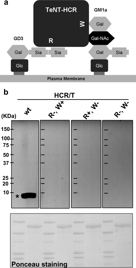
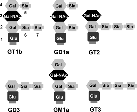
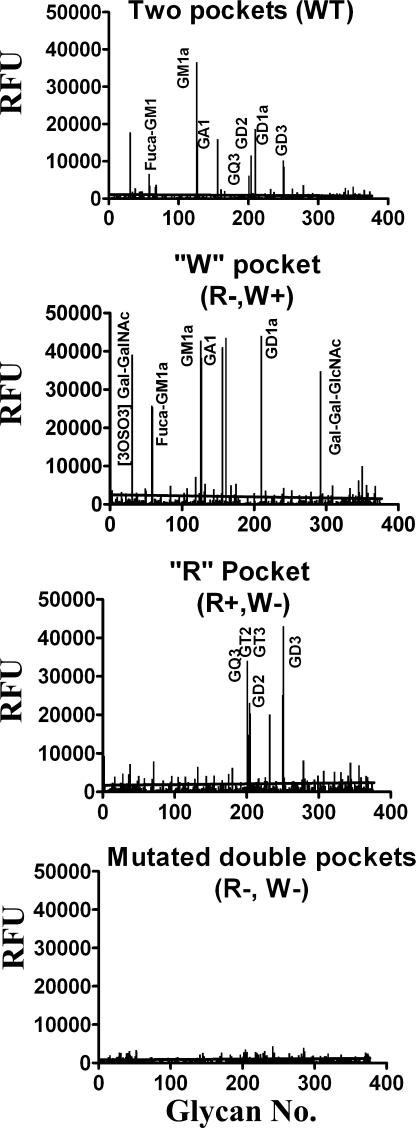
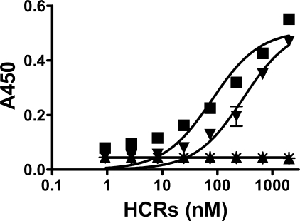
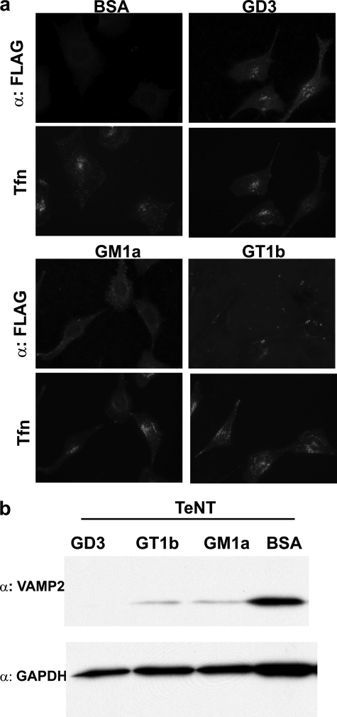
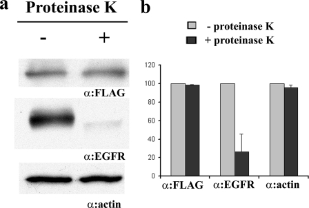
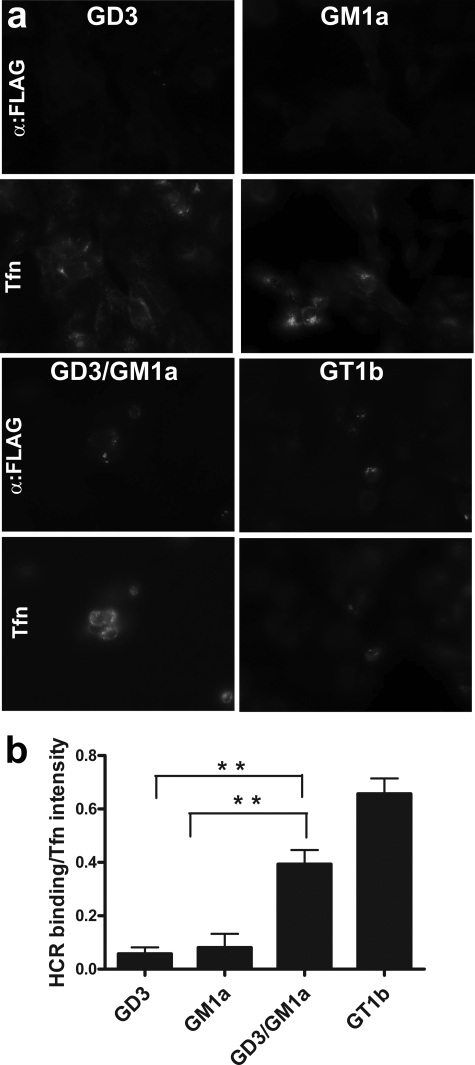
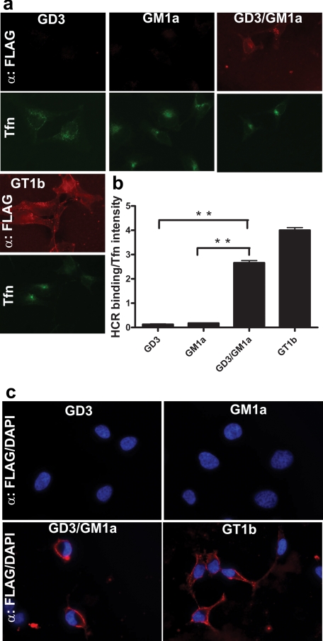
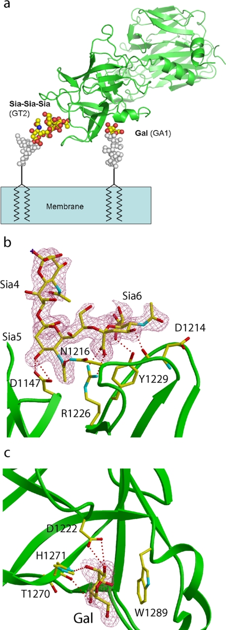
Tetanus neurotoxin (TeNT) is an exotoxin produced by Clostridium tetani that causes paralytic death to hundreds of thousands of humans annually. TeNT cleaves vesicle-associated membrane protein-2, which inhibits neurotransmitter release in the central nervous system to elicit spastic paralysis, but the molecular basis for TeNT entry into neurons remains unclear. TeNT is a approximately 150-kDa protein that has AB structure-function properties; the A domain is a zinc metalloprotease, and the B domain encodes a translocation domain and C-terminal receptor-binding domain (HCR/T). Earlier studies showed that HCR/T bound gangliosides via two carbohydrate-binding sites, termed the lactose-binding site (the "W" pocket) and the sialic acid-binding site (the "R" pocket). Here we report that TeNT high affinity binding to neurons is mediated solely by gangliosides. Glycan array and solid phase binding analyses identified gangliosides that bound exclusively to either the W pocket or the R pocket of TeNT; GM1a bound to the W pocket, and GD3 bound to the R pocket. Using these gangliosides and mutated forms of HCR/T that lacked one or both carbohydrate-binding pocket, gangliosides binding to both of the W and R pockets were shown to be necessary for high affinity binding to neuronal and non-neuronal cells. The crystal structure of a ternary complex of HCR/T with sugar components of two gangliosides bound to the W and R supported the binding of gangliosides to both carbohydrate pockets. These data show that gangliosides are functional dual receptors for TeNT.


Figures

FIGURE 1.
FIGURE 1.
Interaction of the HCR domain of TeNT with its putative cellular receptor. a, HCR/T has two ganglioside-binding sites. The W pocket binds to the terminal GalNAc-Gal of the ganglioside (illustrated by GM1a). The R pocket binds to the di-sialic acid of the ganglioside (illustrated by GD3). b, alternating lanes of molecular mass marker proteins and cortical neuron lysates were separated by SDS-PAGE and transferred to a polyvinylidene difluoride membrane. The membrane was stained for protein with Ponceau S (bottom panel), and then the membrane strips were incubated with 10 nm of the indicated HCR/T (HCR/T wild type (wt), HCR/T (R+, W−), HCR/T (R−, W+), or HCR/T (R−, W−)) followed by HRP-conjugated α-FLAG antibody. The bands were visualized with SuperSignal; exposed film is shown (upper panel). The asterisk denotes the position of purified gangliosides resolved under identical conditions. Migration of the molecular mass marker proteins is indicated (kDa) in the left-most lane in the upper panel.
FIGURE 2.
FIGURE 2.
Schematic of gangliosides described in the study. Gangliosides are composed of ceramide (not shown, but located at the bottom of each structure) linked by a glycosidic bond to an oligosaccharide chain containing hexoses and N-acetylneuraminic acid(s) (sialic acid). Sia, sialic acid; Glu, glucose; Gal, galactose; Gal-NAc, N-acetylgalactosamine.
FIGURE 3.
FIGURE 3.
Glycan array analysis of wild type and mutated HCR/T. Alexa Fluor 488-labeled 200 μg/ml HCR/T, HCR/T (R−, W+), HCR/T (R+, W−), and 100 μg/ml HCR/T (R−, W−) were screened on a protein-glycan array version 3.1 by the Consortium for Functional Glycomics. Glycan array version 3.1 contained 377 derivatives, and HCR/T mainly interacts with ganglioside derivatives that are shown on the graph. The complete data sets of these analyses are available at the Consortium for Functional Glycomics website.
FIGURE 4.
FIGURE 4.
Binding of wild type and mutated HCR/T domains to ganglioside GD3. Immobilized ganglioside GD3 (250 ng/well) was incubated with the indicated HCR domains and assayed for HCR binding as described previously (15). The data were plotted using GraphPad Prism version 5 (GraphPad Software Inc.). ■, HCR/T wild type; ▾, HCR/T (R+, W−); ▴, HCR/T (R−, W+); *, HCR/T (R−, W−).
FIGURE 5.
FIGURE 5.
Ganglioside loading of PC12 cells enhances the uptake of TeNT. PC12 cells were loaded with the indicated ganglioside. a, cells were washed and incubated with 100 nm HCR/T and 10 μg/ml of Alexa Fluor 488-Transferrin (Tfn) for 30 min at 37 °C. HCR/T was visualized by immunofluorescence using a mouse α-FLAG antibody followed by Alexa-coupled secondary IgG. b, cells were incubated with 30 nm TeNT for an additional 48 h. VAMP2 cleavage was visualized by Western blotting, using a mouse α-Syb2 antibody, which recognizes only full-length VAMP2. BSA, bovine serum albumin.
FIGURE 6.
FIGURE 6.
Proteinase K-independent binding of HCR/T on PC12 cells. a, PC12 cells were loaded with 100 μg/ml GT1b for 24 h and treated with or without proteinase K for 30 min at 4 °C. The cells were washed and incubated with 100 nm HCR/T for 1 h at 4 °C. The cells were washed and lysed with radioimmune precipitation assay buffer. HCR/T binding was visualized by Western blot using HRP-conjugated anti-3×FLAG antibody. As control, the extracellular protein epidermal growth factor receptor (EGFR) was visualized by α-epidermal growth factor receptor antibody, and the intracellular protein actin was visualized by α-β actin antibody. b, two independent experiments were quantified using Alpha Innotech Fluorchem system. Immunoblots of the cells treated with proteinase K were presented as a relative level compared with the immunoblots of cells not treated with proteinase K, which were set at 100. The data were presented as a mean with standard error.
FIGURE 7.
FIGURE 7.
Gangliosides reconstitute entry of HCR/T into PC12 cells treated with the glycosphingolipids synthesis inhibitor PPMP. a, PC12 cells were incubated with 25 μm PPMP for 48 h and then loaded with the indicated ganglioside(s) in the presence of PPMP for an additional 24 h. The cells were washed and incubated with 100 nm HCR/T and 10 μg/ml Alexa Fluor 488-Transferrin (Tfn) at 37 °C. Cell-associated HCR was visualized as described above. b, fluorescence was quantified in 20 random fields, averaged, and presented as a fluorescence intensity ratio of bound HCR/Transferrin with standard deviation. Background fluorescence intensities in the absence of exogenous ganglioside loading were subtracted from the ganglioside treated groups. Two-tailed Student's t test, p values <0.05 at the 95% confidence level are indicated by (**) for lined analyses.
FIGURE 8.
FIGURE 8.
Ganglioside-dependent binding of HCR/T to non-neuronal (HeLa) cells. HeLa cells were loaded with the indicated ganglioside. a and c, the cells were washed and incubated with 100 nm HCR/T and 10 μg/ml of Alexa 488-Transferrin (Tfn) either at 37 °C (a) or 4 °C (c). HCR/T was visualized as described above. b, HCR/T fluorescence from a was normalized to transferrin binding, quantified, and processed as described in the legend to Fig. 7 (n = 10).
FIGURE 9.
FIGURE 9.
Structure of HCR/T in complex with ganglioside GT2 and Lactose. a, proposed binding mode of HCR/T on the membrane surface. The structures of gangliosides GT2 and GA1 were modeled onto the complex of HCR/T using the coordinates of HCR/T bound to a GT2 analog and lactose. The HCR is colored green, GT2 analog and galactose are shown in atom coloring, and modeled sugar moieties are shown in gray. b, electron density map (2FoFc map contoured at 1 σ level) around GT2, overlaid with the final refined model (atom coloring, GT2; green, HCR/T). HCR/T residues binding to GT2 are shown in atom coloring. c, electron density map (2FoFc contoured at 1 σ) around galactose, overlaid with the final refined structure (atom coloring, galactose; green, HCR/T). HCR/T residues binding to galactose are shown in atom coloring.

References

    1. Pappas G., Kiriaze I. J., Falagas M. E. (2008) Int. J. Infect. Dis. 12, 347–350 - PubMed
    1. Schiavo G., Benfenati F., Poulain B., Rossetto O., Polverino de Laureto P., DasGupta B. R., Montecucco C. (1992) Nature 359, 832–835 - PubMed
    1. Bilous J., Eggers R., Gasse F., Jarrett S., Lydon P., Magan A., Okwo-Bele J. M., Salama P., Vandelaer J., Villeneuve P., Wolfson L. (2006) Lancet 367, 1464–1466 - PubMed
    1. Schiavo G., Papini E., Genna G., Montecucco C. (1990) Infect. Immun. 58, 4136–4141 - PMC - PubMed
    1. Burton R. M., Balfour Y. M. (1962) Biochem. Pharmacol. 11, 974–976 - PubMed

Publication types

MeSH terms

LinkOut - more resources