. 2009 Jul 15;4(7):e6230.
doi: 10.1371/journal.pone.0006230.

Eyes wide shut: amygdala mediates eyes-closed effect on emotional experience with music

Affiliations

Eyes wide shut: amygdala mediates eyes-closed effect on emotional experience with music

Yulia Lerner et al. PLoS One. .

Abstract

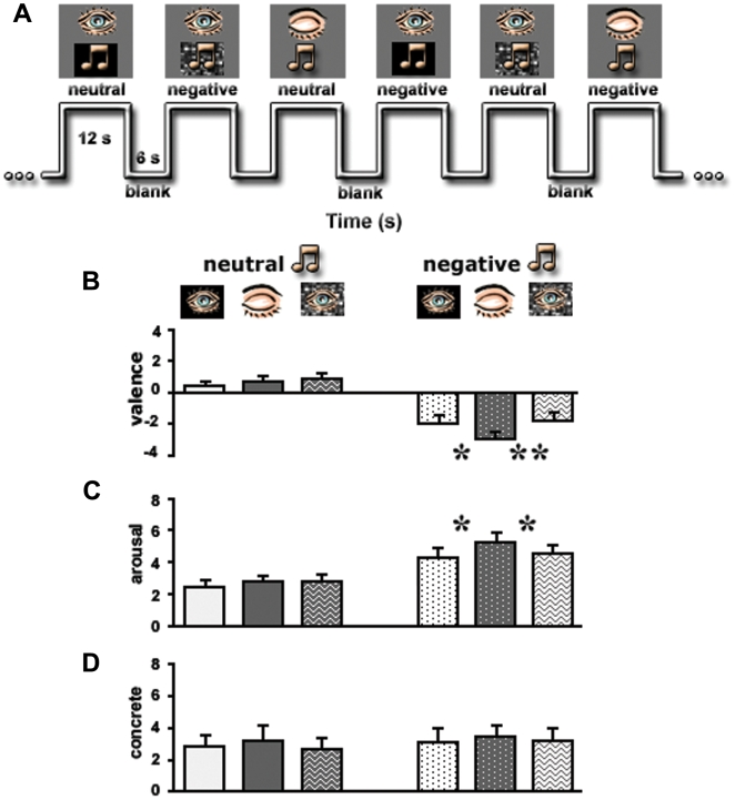
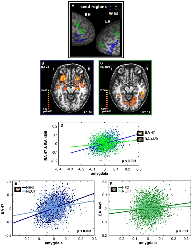
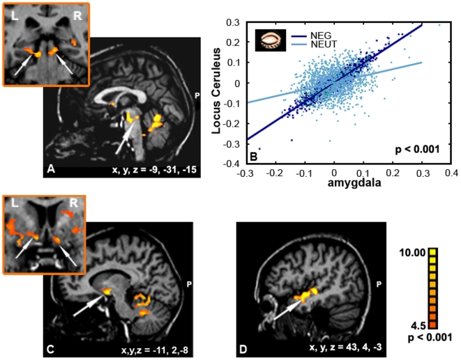
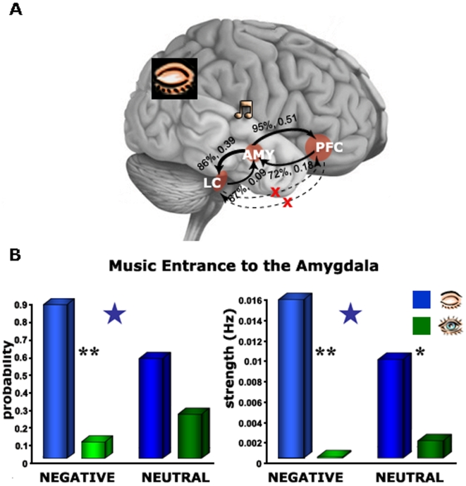
The perceived emotional value of stimuli and, as a consequence the subjective emotional experience with them, can be affected by context-dependent styles of processing. Therefore, the investigation of the neural correlates of emotional experience requires accounting for such a variable, a matter of an experimental challenge. Closing the eyes affects the style of attending to auditory stimuli by modifying the perceptual relationship with the environment without changing the stimulus itself. In the current study, we used fMRI to characterize the neural mediators of such modification on the experience of emotionality in music. We assumed that closed eyes position will reveal interplay between different levels of neural processing of emotions. More specifically, we focused on the amygdala as a central node of the limbic system and on its co-activation with the Locus Ceruleus (LC) and Ventral Prefrontal Cortex (VPFC); regions involved in processing of, respectively, 'low', visceral-, and 'high', cognitive-related, values of emotional stimuli. Fifteen healthy subjects listened to negative and neutral music excerpts with eyes closed or open. As expected, behavioral results showed that closing the eyes while listening to emotional music resulted in enhanced rating of emotionality, specifically of negative music. In correspondence, fMRI results showed greater activation in the amygdala when subjects listened to the emotional music with eyes closed relative to eyes open. More so, by using voxel-based correlation and a dynamic causal model analyses we demonstrated that increased amygdala activation to negative music with eyes closed led to increased activations in the LC and VPFC. This finding supports a system-based model of perceived emotionality in which the amygdala has a central role in mediating the effect of context-based processing style by recruiting neural operations involved in both visceral (i.e. 'low') and cognitive (i.e. 'high') related processes of emotions.


Conflict of interest statement

Competing Interests: The authors have declared that no competing interests exist.

Figures

Figure 1
Figure 1. Experimental design and behavioral results.
(A) A segment from the time axis of the experiment. (B)–(D) Results of psychophysical tests conducted outside the magnet on the same subjects who participated in the fMRI study and using the same stimuli. Evaluation of the emotional valence (B), arousal (C) and abstraction (D) levels of the clips are presented. Stimuli types (x-axis) are identified in the apertures above the graphs. Note a significant difference only for negative clips presented with eyes closed. Y-axes represent emotional dimensions. *, p<0.05; **, p<0.01. Error bars, SEM.
Figure 2
Figure 2. Differences in brain activation with eyes closed, eyes open and activation profiles of ROI.
Functional averaged activation maps (N = 12, p<0.05, random effect) show the cortical activity evoked by musical clips (negative and neutral) presented with eyes closed (blue) and open (green). The maps are superimposed on the left (LH) and right (RH) unfolded hemispheres shown in the lateral view. Quantitative analysis of the activation levels is shown for open (green patches on the maps) and closed (blue patches on the maps) eyes. While the BA 47 (‘blue-framed’ bars, blue circle on the map) exhibited highly preferential activation for eyes closed, the BA 46/9 (‘green-framed’ bars, green circle on the map) demonstrated highly preferential activation for eyes open. STS – superior temporal sulcus, IPS – intraparietal sulcus, PCS – post-central sulcus, LS – lateral sulcus, IFS – inferior frontal sulcus. Error bars, SEM. *, p<0.05.
Figure 3
Figure 3. The effect of eyes closed on the amygdala.
Average activation patterns (N = 12, p<0.05, random effect GLM analysis) revealed by the contrast ‘eyes closed > eyes open’ and superimposed on the coronal and transversal views. Significant activation was found in the amygdala/anterior hippocampus complex (orange arrows) for the negative clips (left panel) but not for the neutral ones (right panel). The color scale indicates significance level. L – left hemisphere, R – right hemisphere, COR – coronal, TRA – transversal.
Figure 4
Figure 4. Sensitivity to emotional clips as reflected in activation profiles.
(A) Average activation levels obtained in the amygdala. Stimulus type (x-axis) is indicated in the apertures above the graphs. The y-axis denotes an fMRI percent signal change relative to blanks. Asterisks denote a significant difference between eye states; p<0.001. Error bars, SEM. (B) ‘Index of eye state’ expressed by formula (close – open)/(close+open) indicates the ratios for negative and neutral stimuli. Asterisk denotes a significant difference between emotions; p<0.05. Error bars, SEM. (C) Relationship of the fMRI data and behavioral performance. Significant correlation (p<0.05) was found only for the amygdala between difference in measure of activation (negative closed – negative open, %SC – % signal change) and difference in the arousal intensity for negative clips. (D) Significant correlation (p<0.05) was found in the amygdala between difference in brain measures (negative closed – neutral closed, %SC – % signal change) and difference in the arousal intensity for eyes closed state.
Figure 5
Figure 5. Correlation analysis for the PFC ROIs.
(A) ‘Seed’ time courses defined separately for eyes open and closed are shown on the parts of unfolded hemispheres. ‘Seed’ time courses were used to compute a voxel-by-voxel fit for the (B) BA 47 and for the (C) BA 46/9. Note a strong connection of the amygdala to the BA 47. The color scale indicates significance level. R – right hemisphere, L – left hemisphere. (D) Correlated activity of the VPFC (BA 47) and amygdala in comparison to the correlated activity of the DLPFC (BA 46/9) and amygdala, corresponding to mental sets of eyes closed and open, respectively. Note a significant difference between correlation coefficients. (E–F) Co-activation analyses respectively to the emotional context in music. A statistical comparison between the co-activation in the negative and neutral conditions was performed for the BA47 (E) and BA 46/9 (F).
Figure 6
Figure 6. Correlation analysis for the amygdala ROI.
An additional network of co-activation was revealed as during closed eyes in negative music. Arrows point to main regions of correlated activity with the ROI: (A) LC, (C) NAcc, and (D) anterior temporal gyrus. (B) Co-activation respectively to the emotional context is depicted. The correlated activity between the amygdala and LC during eyes closed was much stronger in the negative emotional condition than in the neutral condition.
Figure 7
Figure 7. DCM – model and connectivity.
(A) directions of the connectivity between the amygdala, PFC and LC during eyes closed. (B) The power of intrinsic connections indicated by probability and strength. Note the most probable and the strongest connection from the amygdala toward the PFC. *, p<0.01, **, p<0.001. Blue stars indicate a significant difference between the valences under closed eyes state (p<0.05).

References

    1. Gilden DL. Cognitive emissions of 1/f noise. Psychol Rev. 2001;108:33–56. - PubMed
    1. Berger H. On the electroencephalogram in man. Archiv fur Psychiatrie und nervenkrankheiten. 1929;87:527–543.
    1. Ben-Simon E, Podlipsky I, Arieli A, Zhdanov A, Hendler T. Never Resting Brain: Simultaneous Representation of Two Alpha Related Processes in Humans. PLoS ONE. 2008;3:e3984. doi:3910.1371/journal.pone.0003984. - PMC - PubMed
    1. James W. What is an Emotion? Mind. 1884;9:188–205.
    1. Damasio A. Feelings of emotion and the self. Ann N Y Acad Sci. 2003;1001:253–261. - PubMed

Publication types