. 2009 Sep-Oct;15(9-10):328-36.
doi: 10.2119/molmed.2009.00016. Epub 2009 Jun 26.

Progesterone with vitamin D affords better neuroprotection against excitotoxicity in cultured cortical neurons than progesterone alone

Affiliations

Progesterone with vitamin D affords better neuroprotection against excitotoxicity in cultured cortical neurons than progesterone alone

Fahim Atif et al. Mol Med. 2009 Sep-Oct.

Abstract

Because the complex heterogeneity of traumatic brain injury (TBI) is believed by many to be a major reason for the failed clinical trials of monotherapies, combining two (or more) drugs with some potentially different mechanisms of action may produce better effects than administering those agents individually. In this study, we investigated whether combinatorial treatment with progesterone (PROG) and 1,25-dihydroxyvitamin D(3) hormone (VDH) would produce better neuroprotection than PROG alone following excitotoxic neuronal injury in vitro. E18 rat primary cortical neurons were pretreated with various concentrations of PROG and VDH separately or in combination for 24 h and then exposed to glutamate (0.5 micromol/L) for the next 24 h. Lactate dehydrogenase (LDH) release and 3-(4,5-dimethylthiazol-2-yl)-2,5-diphenyltetrazolium bromide (MTT) reduction assays were used to measure cell death. Both PROG and VDH significantly (P < 0.001) reduced neuronal loss when tested independently. Primary cortical cultures treated with VDH exhibited a U-shaped concentration-response curve. PROG at 20 micromol/L and VDH at 100 nmol/L concentrations were the most neuroprotective. When the drugs were combined, the "best" doses of PROG (20 micromol/L) and VDH (100 nmol/L), used individually, did not show substantial efficacy; rather, the lower dose of VDH (20 nmol/L) was most effective when used in combination with PROG (P < 0.01). We also examined the effect of combinatorial treatment on mitogen-activated protein kinase (MAPK) activation as a potential neuroprotective mechanism and observed that PROG and VDH activated MAPK alone and in combination. Interestingly, the best combination dose of PROG and VDH (20 micromol/L and 20 nmol/L, respectively), as observed in cell death assays (LDH and MTT), resulted in increased MAPK activation compared with either the most neuroprotective concentration of individual PROG (20 micromol/L) and VDH (100 nmol/L) or the combination of these individual best doses. Such interactions must be considered in planning individualized combinatorial therapies. In conclusion, the findings of the present study can be taken to suggest that VDH warrants study as a potential partner for combination therapy with PROG.


Figures

Figure 1
Figure 1
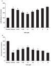
Effect of PROG on glutamate-induced LDH release (A) and MTT reduction (B) in rat primary cortical neurons. Primary cells were pretreated with different concentrations of PROG for 24 h and subsequently exposed to glutamate (0.5 μmol/L) for 24 h. PROG was present in the culture medium during the glutamate exposure. The values are expressed as mean ± SEM of four experiments. Significant difference: #P < 0.001 compared with control; *P < 0.001 compared with vehicle.
Figure 2
Figure 2
Effect of VDH on glutamate-induced LDH release (A) and MTT reduction (B) in rat primary cortical neurons. Primary cells were pretreated with different concentrations of VDH for 24 h and subsequently exposed to glutamate (0.5 μmol/L) for 24 h. VDH was present in the culture medium during the glutamate exposure. The values are expressed as mean ± SEM of four experiments. Significant difference: #P < 0.001 compared with control; *P < 0.001 compared with vehicle.
Figure 3
Figure 3
Effect of combinatorial treatment of PROG and VDH on glutamate-induced LDH release (A) and MTT reduction (B) in rat primary cortical neurons. Primary cells were pre-treated with either best concentrations of PROG and VDH or their combination for 24 h and subsequently exposed to glutamate (0.5 μmol/L) for 24 h. Drugs were present in the culture medium during the glutamate exposure. The values are expressed as mean ± SEM of three experiments. Significant difference: #P < 0.001 compared with control; *P < 0.001 compared with vehicle.
Figure 4
Figure 4
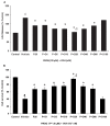
Effect of combinatorial treatment of PROG and VDH on glutamate-induced LDH release (A) and MTT reduction (B) in rat primary cortical neurons. Primary cells were pre-treated with different combinations of PROG and VDH for 24 h and subsequently exposed to glutamate (0.5 μmol/L) for 24 h. Drugs were present in the culture medium during the glutamate exposure. The values are expressed as mean ± SEM of four experiments. Significant difference: #P < 0.001 compared with control, *P < 0.001 compared with vehicle, and §P < 0.01 compared with P20 group.
Figure 5
Figure 5
Changes in the morphology of neurons treated with VDH or PROG. Control group shows neuritic processes characteristic of neurons in vitro. The glutamate (0.5 μmol/L for 24 h) group shows the loss of neuronal processes and number of neurons. PROG (20 μmol/L) and VDH (100 nmol/L) alone rescued neurons independently, but this neuroprotective effect was more pronounced when neurons were exposed to their combination, that is, PROG (20 μmol/L) and VDH (20 nmol/L).
Figure 6
Figure 6
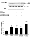
Effect of PROG and VDH exposure on the activation of MAPK in primary cortical neurons. Cells were exposed to hormones either separately or in various combinations for 30 min. Cells were lysed after incubation, and lysates were separated on 12.5% SDS gel and transferred onto polyvinylidene difluoride membrane. Membrane was probed with either phospho-ERK1/2 or total ERK1/2 protein. Phospho-ERK1/2 data were normalized with total ERK1/2 protein. Data were analyzed using ANOVA and the Neuman–Keuls test. Values are expressed as SEM of three independent experiments. Significant difference: *P < 0.05 compared with control; P < 0.05 compared with PROG (20 μmol/L) and VDH (20 nmol/L) groups. Values in parentheses represent fold increase in MAPK over control values.

References

    1. Doppenberg EM, Choi SC, Bullock R. Clinical trials in traumatic brain injury: lessons for the future. J Neurosurg Anesthesiol. 2004;16:87–94. - PubMed
    1. Sidaros A, et al. Long-term global and regional brain volume changes following severe traumatic brain injury: a longitudinal study with clinical correlates. Neuroimage. 2009;44:1–8. - PubMed
    1. Behan LA, Agha A. Endocrine consequences of adult traumatic brain injury. Horm Res. 2007;68(Suppl 5):18–21. - PubMed
    1. Margulies S, Hicks R the Combination Therapies for Traumatic Brain Injury Workshop Leaders. Combination therapies for traumatic brain injury: prospective considerations. J Neuro-trauma. 2009;26:925–39. - PMC - PubMed
    1. Wright DW, et al. ProTECT: a randomized clinical trial of progesterone for acute traumatic brain injury. Ann Emerg Med. 2007;49:391–402. - PubMed

Publication types

MeSH terms