

Severe burn-induced endoplasmic reticulum stress and hepatic damage in mice
- PMID: 19603103
- PMCID: PMC2710291
- DOI: 10.2119/molmed.2009.00048
Severe burn-induced endoplasmic reticulum stress and hepatic damage in mice
Abstract
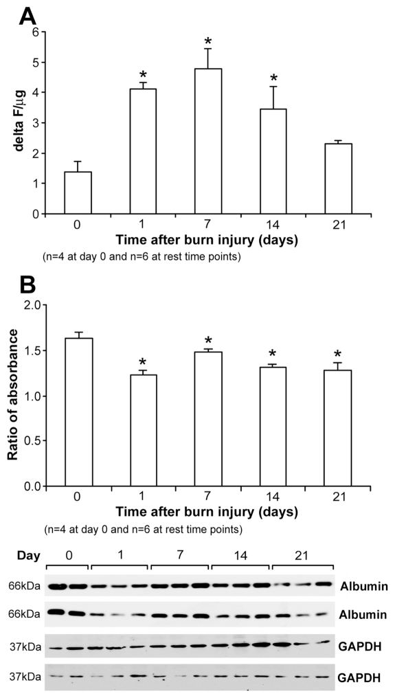
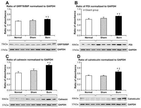
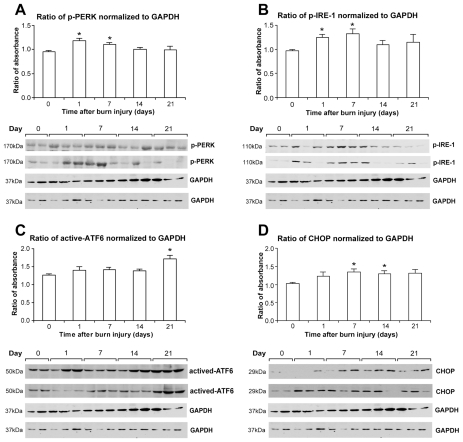
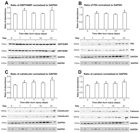
Severe burn injury results in liver dysfunction and damage, with subsequent metabolic derangements contributing to patient morbidity and mortality. On a cellular level, significant postburn hepatocyte apoptosis occurs and likely contributes to liver dysfunction. However, the underlying mechanisms of hepatocyte apoptosis are poorly understood. The endoplasmic reticulum (ER) stress response/unfolded protein response (UPR) pathway can lead to hepatocyte apoptosis under conditions of liver dysfunction. Thus, we hypothesized that ER stress/UPR may mediate hepatic dysfunction in response to burn injury. We investigated the temporal activation of hepatic ER stress in mice after a severe burn injury. Mice received a scald burn over 35% of their body surface and were killed at 1, 7, 14, and 21 d postburn. We found that severe burn induces hepatocyte apoptosis as indicated by increased caspase-3 activity (P < 0.05). Serum albumin levels decreased postburn and remained lowered for up to 21 d, indicating that constitutive secretory protein synthesis was reduced. Significantly, upregulation of the ER stress markers glucose-related protein 78 (GRP78)/BIP, protein disulfide isomerase (PDI), p-protein kinase R-like endoplasmic reticulum kinase (p-PERK), and inositol-requiring enzyme 1alpha (IRE-1alpha) were found beginning 1 d postburn (P < 0.05) and persisted up to 21 d postburn (P < 0.05). Hepatic ER stress induced by burn injury was associated with compensatory upregulation of the calcium chaperone/storage proteins calnexin and calreticulin (P < 0.05), suggesting that ER calcium store depletion was the primary trigger for induction of the ER stress response. In summary, thermal injury in mice causes long-term adaptive and deleterious hepatic function alterations characterized by significant upregulation of the ER stress response.
Figures
Similar articles
-
Insulin protects against hepatic damage postburn.Mol Med. 2011 May-Jun;17(5-6):516-22. doi: 10.2119/molmed.2010.00166. Epub 2011 Jan 19. Mol Med. 2011. PMID: 21267509 Free PMC article.
-
Calcium and ER stress mediate hepatic apoptosis after burn injury.J Cell Mol Med. 2009 Aug;13(8B):1857-65. doi: 10.1111/j.1582-4934.2009.00644.x. J Cell Mol Med. 2009. PMID: 20141609 Free PMC article.
-
Hepatic autophagy after severe burn in response to endoplasmic reticulum stress.J Surg Res. 2014 Mar;187(1):128-33. doi: 10.1016/j.jss.2013.09.042. Epub 2013 Oct 2. J Surg Res. 2014. PMID: 24209807 Free PMC article.
-
The critical roles of endoplasmic reticulum chaperones and unfolded protein response in tumorigenesis and anticancer therapies.Oncogene. 2013 Feb 14;32(7):805-18. doi: 10.1038/onc.2012.130. Epub 2012 Apr 16. Oncogene. 2013. PMID: 22508478 Free PMC article. Review.
-
Endoplasmic reticulum chaperones tweak the mitochondrial calcium rheostat to control metabolism and cell death.Cell Calcium. 2018 Mar;70:64-75. doi: 10.1016/j.ceca.2017.05.015. Epub 2017 May 31. Cell Calcium. 2018. PMID: 28619231 Review.
Cited by
-
Severe Burn-Induced Intestinal Epithelial Barrier Dysfunction Is Associated With Endoplasmic Reticulum Stress and Autophagy in Mice.Front Physiol. 2018 Apr 23;9:441. doi: 10.3389/fphys.2018.00441. eCollection 2018. Front Physiol. 2018. PMID: 29740349 Free PMC article.
-
Beneficial Effects of Systemically Administered Human Muse Cells in Adriamycin Nephropathy.J Am Soc Nephrol. 2017 Oct;28(10):2946-2960. doi: 10.1681/ASN.2016070775. Epub 2017 Jul 3. J Am Soc Nephrol. 2017. PMID: 28674043 Free PMC article.
-
NLRP3 knockout enhances immune infiltration and inflammatory responses and improves survival in a burn sepsis model.Immunology. 2022 Feb;165(2):195-205. doi: 10.1111/imm.13427. Epub 2021 Nov 24. Immunology. 2022. PMID: 34773253 Free PMC article.
-
Plasmin drives burn-induced systemic inflammatory response syndrome.JCI Insight. 2021 Dec 8;6(23):e154439. doi: 10.1172/jci.insight.154439. JCI Insight. 2021. PMID: 34877937 Free PMC article.
-
β-Adrenergic blockade attenuates adverse adipose tissue responses after burn.J Mol Med (Berl). 2024 Oct;102(10):1245-1254. doi: 10.1007/s00109-024-02478-w. Epub 2024 Aug 15. J Mol Med (Berl). 2024. PMID: 39145814
References
-
- Herndon DN, Hart DW, Wolf SE, Chinkes DL, Wolfe RR. Reversal of catabolism by beta-blockade after severe burns. N Engl J Med. 2001;345:1223–9. - PubMed
-
- Pereira C, Murphy K, Herndon D. Outcome measures in burn care: is mortality dead? Burns. 2004;30:761–71. - PubMed
-
- Brusselaers N, Monstrey SJ, Vandijck DM, Blot SI. Prediction of morbidity and mortality on admission to a burn unit. Plast Reconstr Surg. 2007;120:360–1. author reply 361. - PubMed
-
- Jeschke MG, Micak RP, Finnerty CC, Herndon DN. Changes in liver function and size after a severe thermal injury. Shock. 2007;28:172–7. - PubMed
-
- Thomas S, Wolf SE, Chinkes DL, Herndon DN. Recovery from the hepatic acute phase response in the severely burned and the effects of long-term growth hormone treatment. Burns. 2004;30:675–9. - PubMed
Publication types
MeSH terms
Substances
Grants and funding
LinkOut - more resources
Full Text Sources
Medical
Research Materials
Miscellaneous
