Review
. 2009 Sep-Oct;15(9-10):337-51.
doi: 10.2119/molmed.2009.00005. Epub 2009 Apr 10.

The hepatic response to thermal injury: is the liver important for postburn outcomes?

Affiliations
Review

The hepatic response to thermal injury: is the liver important for postburn outcomes?

Marc G Jeschke. Mol Med. 2009 Sep-Oct.

Abstract

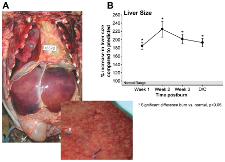
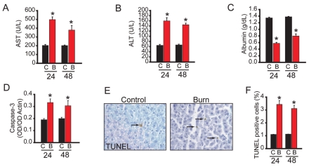
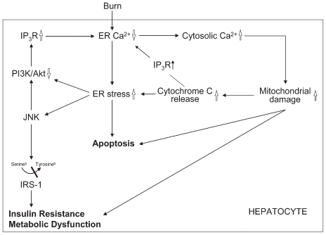
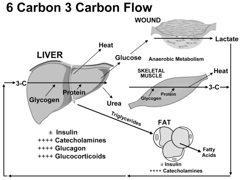
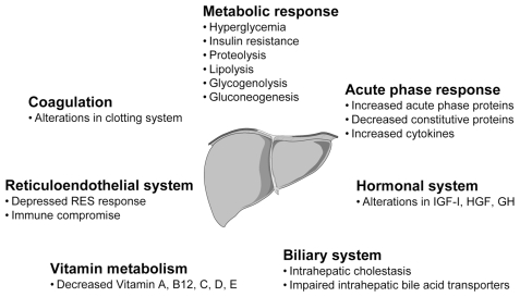
Thermal injury produces a profound hypermetabolic and hypercatabolic stress response characterized by increased endogenous glucose production via gluconeogenesis and glycogenolysis, lipolysis, and proteolysis. The liver is the central body organ involved in these metabolic responses. It is suggested that the liver, with its metabolic, inflammatory, immune, and acute phase functions, plays a pivotal role in patient survival and recovery by modulating multiple pathways following thermal injury. Studies have evaluated the role and function of the liver during the postburn response and showed that liver integrity and function are essential for survival, and that hepatic acute phase proteins are strong predictors for postburn survival. This review discusses these studies and delineates the pivotal role of the liver in patients following severe thermal injury.


Figures

Figure 1
Figure 1
(A) Massive hepatomegaly (upper Figure 1A) and hepatic fatty infiltration (lower Figure 1A) of a burn victim at autopsy. (B) Liver size increases throughout acute hospitalization by over 200% in 242 surviving burn patients. Reprinted with permission from Jeshcke MG, et al. (2008) Pathophysiologic Response to Severe Burn Injury. Ann Surg. 248:387–401.
Figure 2
Figure 2
Hepatic dysfunction of the rat burn model mimics the postburn human disease state. (A) Serum aspartate amino transferase (AST) 24 and 48 h after thermal injury. (B) Serum alanine amino transferase (ALT) 24 and 48 h after thermal injury. (C) Serum albumin 24 and 48 h after thermal injury. (D) Caspase-3 activity in liver lysates as determined by successive Western blotting with active caspase-3 and actin antibodies. The data is expressed as a ratio of the two band intensities. (E) TUNEL staining of a liver section before (Control) and 24 h after thermal injury (Burn). (F) Quantified TUNEL positive cells 24 and 48 h after thermal injury. Time after injury in h is indicated. C, control; B, burn. Data presented are mean ± SEM. *P < 0.05 (Burned animals n = 8 and controls n = 4 per time point). *Significant difference between burn and control, P < 0.05. Reprinted with permission from Jeschke MG, et al. (2008) Calcium and ER Stress Mediate Hepatic Apoptosis after Burn Injury. J Cell Mol Med. Accepted for publication. [Epub ahead of print 2009 Jan 14].
Figure 3
Figure 3
Schematic of suggested pathways involved in the hepatic response postburn. Thermal injury leads to gross alterations in ER calcium with increased cytosolic calcium concentration. Increased cytosolic calcium induces mitochondrial damage which releases cytochrome c. Cytochrome c increases the existing ER stress/UPR but also binds to the IP3R augmenting the depletion of ER calcium stores. ER stress/UPR leads to cell apoptosis and activation of JNK which phosphorylates the 612 serine of IRS-1, which blocks phosphorylation of tyrosine IRS-1. ER stress/UPR also impairs the prosurvival PI3K/Akt signaling which results in an increased activation of the IP3R increasing ER stress/UPR.
Figure 4
Figure 4
Metabolic changes postburn with the liver playing an essential role.
Figure 5
Figure 5
Overview of hepatic functions and responses.

References

    1. Bringham PA, McLoughlin E. Burn incidence and medical care use in the United States: estimates, trends and data sources. J Burn Care Rehabil. 1996;17:95–107. - PubMed
    1. World Health Organization. Facts about injuries: Burns. Geneva: WHO, Injuries & Violence Prevention, Non-Communicable Diseases and Mental Health; [place unknown]: International Society for Burn Injuries; (WHO), Injuries & Violence Prevention, Non-Communicable Diseases and Mental Health; p. 5. No date. Listed as a 2004 fact sheet at: http://www.who.int/violence_injury_prevention/publications/other_injury/....
    1. Palmieri TL, Greenhalgh DG. Topical treatment of pediatric patients with burns: a practical guide. Am J Clin Dermatol. 2002;3:529–34. - PubMed
    1. Nelson KJ, Beierle EA. Exhaust system burn injuries in children. J Pediatr Surg. 2005;40:E43–6. - PubMed
    1. Foglia RP, Moushey R, Meadows L, Seigel J, Smith M. Evolving treatment in a decade of pediatric burn care. J Pediatr Surg. 2004;39:957–60. - PubMed

Publication types