. 2009;13(4):R118.
doi: 10.1186/cc7968. Epub 2009 Jul 15.

The change and effect of endothelial progenitor cells in pig with multiple organ dysfunction syndromes

Affiliations

The change and effect of endothelial progenitor cells in pig with multiple organ dysfunction syndromes

Tian Hang Luo et al. Crit Care. 2009.

Abstract

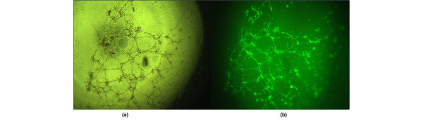
Introduction: The dysfunction and decrease of endothelial progenitor cells (EPCs) may play a very important role in the initiation of organ dysfunction caused by trauma or severe sepsis. We aim to measure the number and function of EPCs in the progression of multiple organ dysfunction syndromes (MODS) caused by severe sepsis, which may help to understand the pathogenesis of MODS by the changing of EPCs.

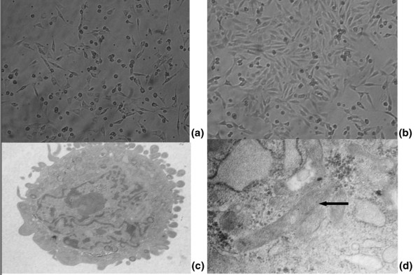
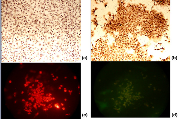
Methods: A total of 40 pigs were randomly divided into two groups, which were subjected to hemorrhagic shock, resuscitation and endotoxemia (experimental group, n = 20) or acted as a control (control group, n = 20). The number and function of EPCs including adhesive, migratory and angiogenesis capacities were analyzed at different times in both groups.

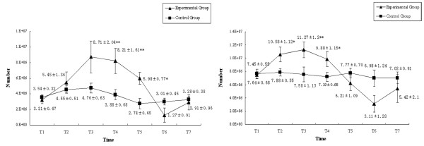
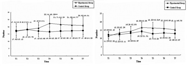
Results: All the animals in the experimental group developed MODS (100%) and 17 of 20 animals (85%) died due to MODS; the incidence of MODS and death of the animals in the control group were 0% (P < 0.01). The number, migratory and adhesive capacities of EPCs decreased sharply in the animals of the experimental group corresponding to the increasing severities of MODS, but the angiogenesis function increased gradually until death. The decrease in function of EPCs preceded the decrease in number of EPCs. The decrease in number and function of EPCs occurred prior to the occurrence of MODS.

Conclusions: For the first time, it was observed that the number and function of EPCs decreased sharply in the progression of MODS and that it was prior to the occurrence of MODS. The decrease in number and function of EPCs may be one of the main pathogenic factors of MODS.


Figures

Figure 1
Figure 1
Number of endothelial progenitor cells during progression of multiple organ dysfunction syndromes. The number of endothelial progenitor cells in (left) peripheral blood and (right) bone marrow. *P < 0.05, **P < 0.01.
Figure 2
Figure 2
Endothelial progenitor cell morphology during ex vivo culture. (a) Mononuclear cells are able to differentiate into spindle cells 48 hours after culture.(b) Colony of endothelial progenitor cells (EPCs) observed after 7 or 8 days of culture. (c) and (d) After 7 days of culture, the ultrastructure of EPCs can be observed by electron microscope. Black arrow, Weible-Palade body that was the characteristic structure of EPCs. Magnification: (a) and (b) ×100; (c) ×5,000; (d) ×30,000.
Figure 3
Figure 3
Characterization of endothelial progenitor cells. At 7 days of culture, immunohistochemical staining revealed that more than 90% of the cultured cells are positive to (a) CD133 and (b) vascular endothelial growth factor receptor-2. Endothelial progenitor cells were incubated with 1,1-dioctadecyl-3,3,3,3-tetramethylindocarbocyanine-labeled acetylated low-density lipoprotein (Dil-Ac-LDL) and stained with Ulex europaeus agglutinin (UEA-1). Fluorescence microscopy illustrates that endothelial progenitor cells are positive for (c) Dil-Ac-LDL and (d) Dil-Ac-LDL and UEA-1. Magnification: ×100.
Figure 4
Figure 4
Migratory and adhesive capacities of endothelial progenitor cells during progression of multiple organ dysfunction syndromes. The (top) migratory capacities and (bottom) adhesive capacities of endothelial progenitor cells in (left) peripheral blood and (right) bone marrow. *P < 0.05, **P < 0.01.
Figure 5
Figure 5
Angiogenic capacity of endothelial progenitor cells during progression of multiple organ dysfunction syndromes. The angiogenic capacity of endothelial progenitor cells in (left) peripheral blood and (right) bone marrow. *P < 0.05, **P < 0.01.
Figure 6
Figure 6
Angiogenesis function of endothelial progenitor cells. Angiogenesis function of endothelial progenitor cells with Calcein added, observed by (a) phase-contrast microscope and (b) fluorescent microscope. Magnification: ×400.

References

    1. Botha AJ, Moore FA, Moore EE, Sauaia A, Banerjee A, Peterson VM. Early neutophil sequestration after injury: a pathogenic mechanism for multiple organ failure. J Trauma. 1995;39:411–417. doi: 10.1097/00005373-199509000-00003. - DOI - PubMed
    1. Ben-Shoshan J, Keren G, George J. Endothelial progenitor cells (EPCs) – new tools for diagnosis and therapy. Harefuah. 2006;145:362–366. - PubMed
    1. Planat-Benard V, Silvestre JS, Cousin B, André M, Nibbelink M, Tamarat R, Clergue M, Manneville C, Saillan-Barreau C, Duriez M, Tedgui A, Levy B, Pénicaud L, Casteilla L. Plasticity of human adipose lineage cells toward endothelial cells: physiological and therapeutic perspectives. Circulation. 2004;109:656–663. doi: 10.1161/01.CIR.0000114522.38265.61. - DOI - PubMed
    1. Asahara T, Murohara T, Sullivan A, Silver M, Zee R van der, Li T, Witzenbichler B, Schatteman G, Isner JM. Isolation of putative endothelial progenitorcells for angiogenesis. Science. 1997;275:964–967. doi: 10.1126/science.275.5302.964. - DOI - PubMed
    1. Yasuhara H, Muto T. Ischemia/reperfusion injury and organ failure. Nippon Geka Gakkai Zasshi. 1998;99:510–517. - PubMed

Publication types