. 2009 Jul 15:10:38.
doi: 10.1186/1471-2172-10-38.

The orphan adapter protein SLY1 as a novel anti-apoptotic protein required for thymocyte development

Affiliations

The orphan adapter protein SLY1 as a novel anti-apoptotic protein required for thymocyte development

Bernhard Reis et al. BMC Immunol. .

Abstract

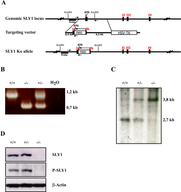
Background: SH3 containing Lymphocyte Protein (SLY1) is a putative adapter protein exclusively expressed in lymphocytes which is involved in antigen receptor induced activation. We previously have generated SLY1Delta/Delta mice harbouring a partial deletion in the N-terminal region of SLY1 which revealed profound immunological defects in T and B cell functions.

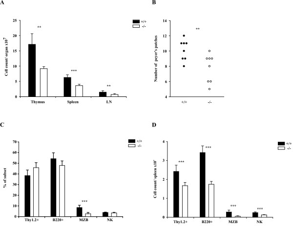
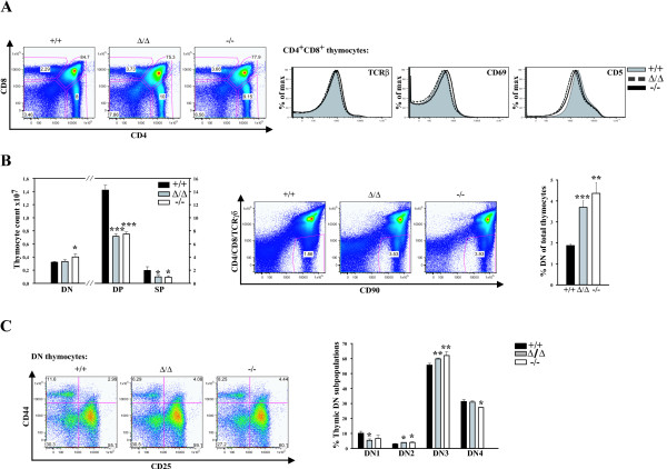
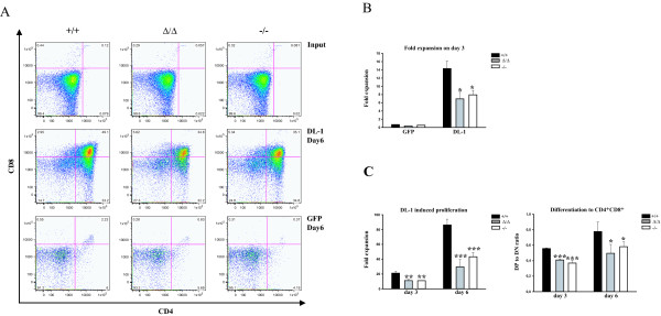
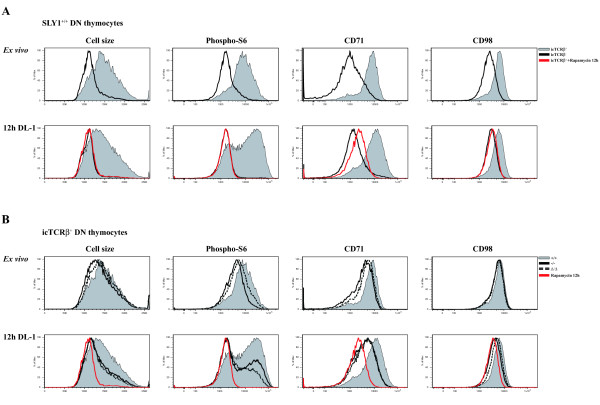
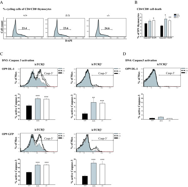
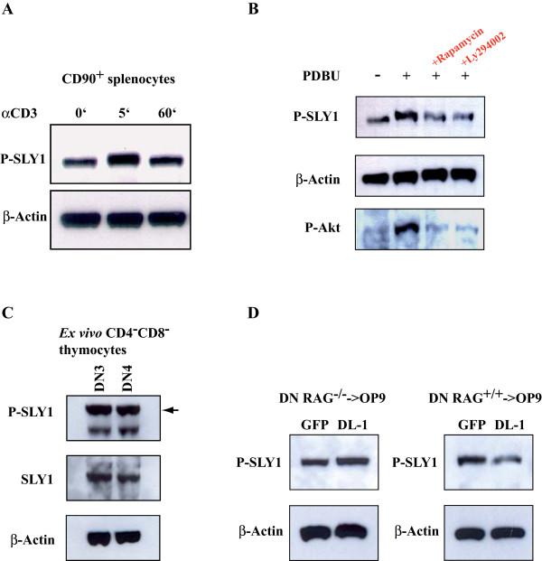
Results: In this study, T cell development in SLY1-/- and SLY1Delta/Delta mice was analysed ex vivo and upon cultivation with the bone marrow stromal cell line OP9. SLY1-deficient thymocytes were compromised in inducing nutrient receptor expression and ribosomal protein S6 phosphorylation, indicating a defect in mTOR complex activation. Furthermore, SLY1 was identified as a novel anti-apoptotic protein required for developmental progression of T cell precursors to the CD4+CD8+ double-positive stage by protecting from premature programmed cell death initiation in developing CD4-CD8- double-negative thymocytes. In addition, SLY1 phosphorylation was differentially regulated upon Notch ligand-mediated stimulation and expression of the preTCR.

Conclusion: Thus, our results suggest a non-redundant role for SLY1 in integrating signals from both receptors in early T cell progenitors in the thymus.


Figures

Figure 1
Figure 1
Generation of SLY1 ko mice. A. Scheme of the sly1 genomic locus, targeting vector and inserted neomycin cassette after homologous recombination. The neomycin cassette is inserted in inverted orientation directly after the starting ATG. B. Screening PCR with genomic tail DNA and primers as described in material and methods. C. Southern blot after digestion of genomic tail DNA with EcoRV and detection with 32P-labeled probe depicted in A. D. Western Blot assessing the successful inactivation of the sly1 gene using two different SLY1-specific antibodies and β-actin as loading control.
Figure 2
Figure 2
Reduced cellularity of lymphoid organs in SLY1-/--mice. A. Mean cell count of thymus, spleen and pooled axial and inguinal lymph nodes was determined (n = 4). B. Reduced number of visible Peyer's patches in SLY1-/--mice. C. FACS analysis of splenic lymphocyte subpopulations. T cells were determined as Thy1.2+, B cells as B220+, marginal zone B cells as CD21highCD23- of B220+, and NK cells as CD3-CD56+ (n = 9). D. Absolute cell counts/spleen (n = 9). **p ≤ 0.01; *** p ≤ 0.001.
Figure 3
Figure 3
Block in thymocyte CD4-CD8- (DN) to CD4+CD8+ (DP) differentiation. A. Total thymocyte CD4- and CD8-expression was determined by FACS analysis (left panel). Histograms (right panel): Surface expression of TCRβ, CD69 and CD5 on gated CD4+CD8+ thymocytes. B. Cell count of DN, DP or CD4- and CD8-single positive (SP) thymocytes (left panel). Gating (middle panel) and percentage of DN thymocytes relative to total thymocytes (right panel). C. DN thymocytes were gated as shown in B and a subset analysis according to expression of CD44 and CD25 was performed. DN1 = CD44+CD25-; DN2 = CD44+CD25+; DN3 = CD44-CD25+; DN4 = CD44-CD25-. Dot plots show representative data of three independent experiments (n = 3). *p ≤ 0.05; **p ≤ 0.001; *** p ≤ 0.0005.
Figure 4
Figure 4
SLY1-targeted DN thymocytes exhibit impaired proliferation and differentiation when cultured on OP9 DL-1 cells. A. CD4 and CD8 FACS profiles of sorted DN thymocytes before cultivation (input, upper row), and after cultivation for six days on OP9 DL-1 (middle row) or on OP9 GFP control cells (lower row). B. DL-1-dependent expansion of DN thymocytes after three days culture. C. Fold expansion on day three and day six (left panel). Differentiation rate (right panel) corresponds to the percentage of DP cells divided by the percentage of remaining DN cells. The expansion rate was determined by counting triplicates and comparing to initial seeding numbers on day 0. Representative data of at least three independently performed experiments is shown. *p ≤ 0.05; **p ≤ 0.005; *** p ≤ 0.001.
Figure 5
Figure 5
mTOR-signalling is diminished in developing SLY1-/- and SLY1Δ/Δ DN thymocytes. Cell size, activation of S6 kinase and expression levels of the nutrient receptors CD71 and CD98 were determined as surrogate markers for mTOR activity in thymocytes. Phosphorylation of ribosomal protein S6 was used as a marker of S6 Kinase activity [25]. A. SLY1+/+ DN thymocytes were analysed ex vivo and after 12 h culture on OP9 DL-1 cells. Thymocytes were further subdivided by gating on preTCR-negative and -positive cells. B. Activation of mTOR in preTCR-expressing SLY1+/+, SLY1-/- and SLY1Δ/Δ thymocytes was analysed ex vivo and after OP9 DL-1 culture in vitro for 12 hours. 20 nM Rapamycin was added as a control during the cultivation period to inhibit mTOR activation. Histograms show representative data of four independent experiments (n = 4).
Figure 6
Figure 6
Increased apoptosis of OP9 cultured DN thymocytes in the absence of functional SLY1 protein. A. Cell cycle analysis of permeabilized DN cells after 2 days of OP9 DL-1 culture using DAPI. B. AnnexinV and DAPI staining of DN thymocytes cultivated for 4 days on OP9 DL-1 cells. C. SLY1-targeting results in increased Caspase-3 activation independent of TCRβ-expression and Notch signalling. Sorted DN3 thymocytes were cultured on OP9 cells for 2 days and then Caspase-3 active fragment in DN cells was analysed dependent on TCRβ-expression. D. Intracellular Caspase-3 activation in DN4 cells is negligible. *p ≤ 0.05; **p ≤ 0.01; *** p ≤ 0.001.
Figure 7
Figure 7
Regulation of SLY1 phosphorylation in thymocytes. A. Phosphorylation of SLY1 at serine27 in peripheral splenocytes. CD90+ MACS-isolated T cells were stimulated with αCD3 (10 μg/ml) for the indicated time and analysed by Western blotting. B. Thymocytes were left unstimulated or incubated for 20' with 20 ng/ml PDBU. Where indicated, cells had been pretreated with Rapamycin (20 nM) or Ly294002 (10 μM) for 1 hour. C. DN3 and DN4 thymocytes were single-cell-sorted according to the expression of CD90, CD25 and CD44, lysed and analysed by Western blotting. D. RAG1-/- or RAG1+/+ DN3 thymocytes were plated over night on OP9 cells. DN thymocytes were then sorted by single-cell flow cytometry, lysed and analysed by Western blotting.

References

    1. Godfrey DI, Kennedy J, Mombaerts P, Tonegawa S, Zlotnik A. Onset of TCR-beta gene rearrangement and role of TCR-beta expression during CD3-CD4-CD8-thymocyte differentiation. J Immunol. 1994;152:4783–4792. - PubMed
    1. Ciofani M, Zuniga-Pflucker JC. The Thymus as an Inductive Site for T Lymphopoiesis. Annu Rev Cell Dev Biol. 2006;23:463–493. doi: 10.1146/annurev.cellbio.23.090506.123547. - DOI - PubMed
    1. von Boehmer H, Aifantis I, Feinberg J, Lechner O, Saint-Ruf C, Walter U, et al. Pleiotropic changes controlled by the pre-T-cell receptor. Curr Opin Immunol. 1999;11:135–142. doi: 10.1016/S0952-7915(99)80024-7. - DOI - PubMed
    1. Strasser A, Huang DC, Vaux DL. The role of the bcl-2/ced-9 gene family in cancer and general implications of defects in cell death control for tumourigenesis and resistance to chemotherapy. Biochim Biophys Acta. 1997;1333:F151–F178. - PubMed
    1. Cory S, Vaux DL, Strasser A, Harris AW, Adams JM. Insights from Bcl-2 and Myc: malignancy involves abrogation of apoptosis as well as sustained proliferation. Cancer Res. 1999;59:1685s–1692s. - PubMed

Publication types

MeSH terms