. 2009 Jul 15:9:41.
doi: 10.1186/1471-213X-9-41.

The caudal regeneration blastema is an accumulation of rapidly proliferating stem cells in the flatworm Macrostomum lignano

Affiliations

The caudal regeneration blastema is an accumulation of rapidly proliferating stem cells in the flatworm Macrostomum lignano

Bernhard Egger et al. BMC Dev Biol. .

Abstract

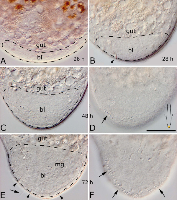
Background: Macrostomum lignano is a small free-living flatworm capable of regenerating all body parts posterior of the pharynx and anterior to the brain. We quantified the cellular composition of the caudal-most body region, the tail plate, and investigated regeneration of the tail plate in vivo and in semithin sections labeled with bromodeoxyuridine, a marker for stem cells (neoblasts) in S-phase.

Results: The tail plate accomodates the male genital apparatus and consists of about 3,100 cells, about half of which are epidermal cells. A distinct regeneration blastema, characterized by a local accumulation of rapidly proliferating neoblasts and consisting of about 420 cells (excluding epidermal cells), was formed 24 hours after amputation. Differentiated cells in the blastema were observed two days after amputation (with about 920 blastema cells), while the male genital apparatus required four to five days for full differentiation. At all time points, mitoses were found within the blastema. At the place of organ differentiation, neoblasts did not replicate or divide. After three days, the blastema was made of about 1420 cells and gradually transformed into organ primordia, while the proliferation rate decreased. The cell number of the tail plate, including about 960 epidermal cells, was restored to 75% at this time point.

Conclusion: Regeneration after artificial amputation of the tail plate of adult specimens of Macrostomum lignano involves wound healing and the formation of a regeneration blastema. Neoblasts undergo extensive proliferation within the blastema. Proliferation patterns of S-phase neoblasts indicate that neoblasts are either determined to follow a specific cell fate not before, but after going through S-phase, or that they can be redetermined after S-phase. In pulse-chase experiments, dispersed distribution of label suggests that S-phase labeled progenitor cells of the male genital apparatus undergo further proliferation before differentiation, in contrast to progenitor cells of epidermal cells. Mitotic activity and proliferation within the blastema is a feature of M. lignano shared with many other regenerating animals.


Figures

Figure 1
Figure 1
Overview of the animal M. lignano. (A) Schematic drawing of M. lignano with indication of the amputation level (red dashed line). Dorsal view. The amputated tail plate is magnified in (C). (B, E, F) Focal interference contrast series through the tail plate of a living specimen. (B) Ventral focus plane showing the male genital opening mo and about 140 duo-gland adhesive systems dg. (C) Schematic drawing of the tail plate. Attached to the male copulatory organ (stylet st) are the vesicula granulorum gv, connected with the seminal vesicle sv and the false seminal vesicle fsv. Prostate glands pg are reaching into the gv and further down the st. Rhabdites rh are located between the epidermal cells. Dg are emerging at the posterior tip of the tail plate. The posterior end of the sac-like gut and cement/shell glands surrounding the female genital opening gsf are at the anterior border of the amputated piece. Main longitudinal nerve cords mnc in blue. (D) Higher magnification of dg. Scale bar is 10 μm. (E) Mnc in focus (band between arrow heads). (F) Mid-level focus plane with the male genital apparatus. Scale bar is 50 μm. (B, E, F) same scale.
Figure 2
Figure 2
The tail plate of M. lignano. (A-C) Electron micrograph of an adult specimen fixed with high pressure freezing. (A) Cross section through the tail plate at the tip of the stylet with representative cell types. Dorsal side is up. (B, C) Details of a more anterior tail plate section than (A). (B) The stylet is surrounded by musculature. The inner epithelial lining is interspersed by muscle cells as well. (C) The tip of a duo-gland adhesive system besides a new epidermis cell in differentiation. ci cilia dg duo-gland adhesive systems, dm dorsoventral muscles, ep multiciliated epidermal cell, gl cyanophil gland cell, mu muscle fibers, ne neoblast, np new epidermal cell, nv nerve, pg prostatic gland, rh rhabdite gland, rm ring muscles, st stylet. (D-G) Fluorescent whole mount stainings of the tail plate. (D) Neoblasts in S-phase labeled with BrdU, (E) epidermal nuclei (inset shows a single lobulated epidermal nucleus) labeled with Hoechst 33342, (F) muscle F-actin stained with phalloidin, (G) GYIRF-amidergic nervous system. Scale bar in (A) is 5 μm, in (B, C) 1 μm, in (D-G) 100 μm.
Figure 3
Figure 3
Early stages of wound closure and healing after amputation. Interference contrast pictures of living specimens.
Figure 4
Figure 4
Interference contrast images of a tail plate blastema of living specimens. 26 (A), 28 (B), 48 (C, D) and 72 (E, F) hours after cutting. The scheme in (D) is indicating the orientation of the animals in all subpanels. (C, D) and (E, F) are different focal planes of the same specimens, respectively. The blastema bl makes up the posterior part of the body and is delimited anteriorly by the gut. 24–28 hours after cutting, the blastema shifts from its ventral position (A) to the posterior end of the body (B). (C) 48 hours after cutting the blastema has grown substantially, and several duo-gland adhesive systems have been rebuilt (arrow in D), which significantly increase in number after 72 hours (F, arrows). At this time, the male genital apparatus mg is being rebuilt with the first tip of the stylet and a small vesicula granulorum to be seen. An arrow points at a long sensory cilium (E). Arrowheads denote rhabdites. Dotted lines indicate blastema extension. Scale bar is 50 μm for all sub-panels.
Figure 5
Figure 5
Semithin sections and whole mounts of 24 hour tail plate blastemas. Schemes are indicating the orientation of the animal in the respective subpanels. (A) Blastema is still located ventrally. Note the mitosis at the anterior end of the blastema (arrowhead). The newly built epidermal layer is characterized through smaller, often less ciliated cells (area above scale bar). A cell migrating to the epidermal layer is shown by the arrow. (B) Blastema is already located caudally. Arrowhead denotes a dorsoventral muscle fiber. (A, B) stained after Heidenhain, (C, E, G) with methylene blue. (C, D, E) BrdU pulse (brown) was applied just before fixation. All three subpanels from the same animal. (C) Complete section, anterior is left. Note the accumulation of labeled neoblasts in the caudal blastema (right). (D) Whole mount and (E) section of the blastema, with accumulation of neoblasts. fa female antrum. (F, G) BrdU pulse chase experiment. Both subpanels are from the same animal. Note the accumulation of labeled cells in the blastema, but the absence of labeled cells in the epidermis. Dotted lines indicate blastema extension. All scale bars are 20 μm, except 50 μm in (C). (D, F) and (E, G) in the same scale.
Figure 6
Figure 6
Semithin sections and whole mounts of 48 hour tail plate blastemas. Schemes are indicating the orientation of the animal in the respective subpanels. (A) At the anterior end (left), the blastema is delimited by the gut. Note the mitoses (arrowheads) within the blastema. An early duo-gland adhesive system, with a cell body close to the epidermis, has been regenerated (double arrowhead). Arrow points at a rhabdite gland. Stained after Heidenhain. (B, C) BrdU pulse. Accumulation of labeled cells in the blastema in whole mount (B) and section (C). Asterisk marks a label-free spot, the presumptive point of origin of the male genital apparatus. (B, C) from the same animal. (D, E) BrdU pulse-chase. (D) Labeled cells are concentrated in the blastema, and also appear in the epidermis (arrowhead). (E) Section of a different animal. Arrowheads in (D, E) point at labeled epidermal cells. Dotted lines indicate blastema extension. (C, E) stained with methylene blue. Scale bars are 20 μm.
Figure 7
Figure 7
Semithin sections and whole mounts of 72 hour tail plate blastemas. Schemes are indicating the orientation of the animal in the respective subpanels. (A-E) Stained after Heidenhain, all pictures from the same series. (A) A regenerated duo-gland adhesive system dg is visible (double arrowhead). Arrowheads denote the regenerated tip of the male genital apparatus mg, consecutive sections showing the composition of this organ in (B-E). (F-H) BrdU pulse, all pictures from the same animal. (F) Whole mount. No accumulation of labeled cells. (G) Section in the area of the regenerating mg (densely blue stained area at the top). No labeled cells (brown) are present in the mg. (H) Section showing a number of labeled cells aside from the mg. (J-L) BrdU pulse-chase. (J) Whole mount, arrowhead points at a labeled epidermal cell. The accumulation of neoblasts in the blastema is evident. (K) Same animal as (J), note the labeled epidermal cells (arrowheads, inset) and the labeled nuclei of the dg (arrow). The regenerating mg is located at the top and shows freckled label in many cells (also see right inset). (L) Horizontal section of a different animal, arrowhead points at the tip of the regenerating mg. (G, H, K, L) stained with methylene blue. Scale bars are 20 μm, (B-E) same scale bar.

References

    1. Ladurner P, Rieger R, Baguñà J. Spatial distribution and differentiation potential of stem cells in hatchlings and adults in the marine platyhelminth Macrostomum sp.: A bromodeoxyuridine analysis. Dev Biol. 2000;226:231–241. - PubMed
    1. Newmark PA, Sánchez Alvarado A. Bromodeoxyuridine specifically labels the regenerative stem cells of planarians. Dev Biol. 2000;220:142–153. - PubMed
    1. Gschwentner R, Ladurner P, Nimeth K, Rieger R. Stem cells in a basal bilaterian. S-phase and mitotic cells in Convolutriloba longifissura (Acoela, Platyhelminthes) Cell Tissue Res. 2001;304:401–408. - PubMed
    1. Egger B, Gschwentner R, Rieger R. Free-living flatworms under the knife: past and present. Dev Genes Evol. 2007;217:89–104. - PMC - PubMed
    1. Paulus T, Müller MCM. Cell proliferation dynamics and morphological differentiation during regeneration in Dorvillea bermudensis (Polychaeta, Dorvilleidae) J Morphol. 2006;267:393–403. - PubMed

Publication types