. 2009 Sep 4;284(36):24035-48.
doi: 10.1074/jbc.M109.014266. Epub 2009 Jul 15.

Biglycan, a danger signal that activates the NLRP3 inflammasome via toll-like and P2X receptors

Affiliations

Biglycan, a danger signal that activates the NLRP3 inflammasome via toll-like and P2X receptors

Andrea Babelova et al. J Biol Chem. .

Abstract

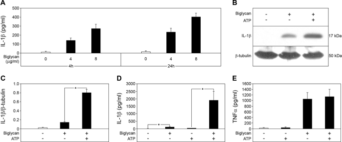
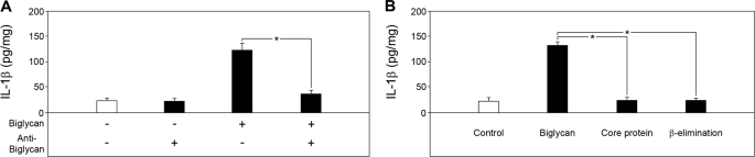
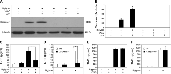
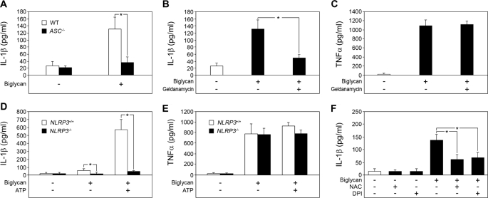
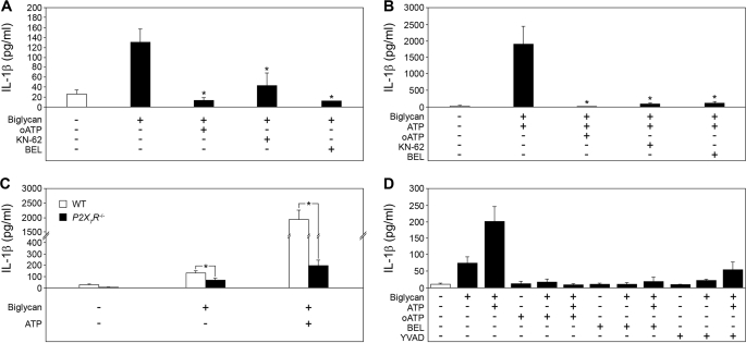
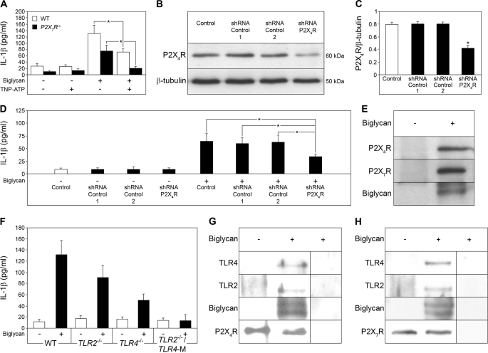
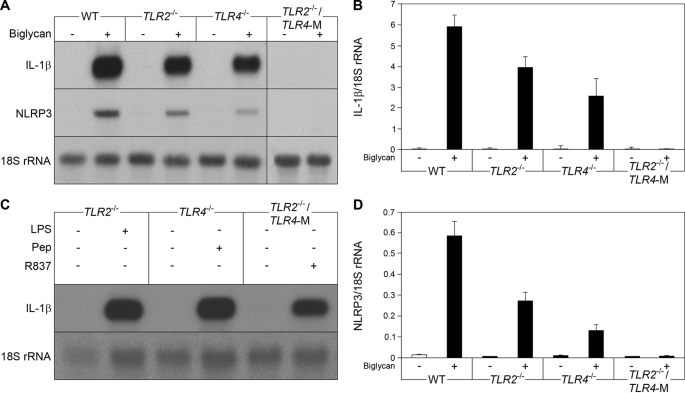
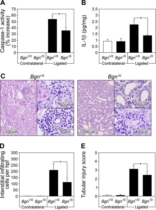
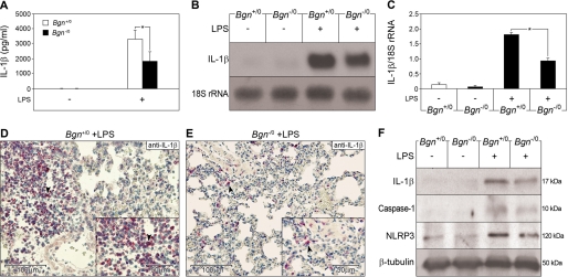
The role of endogenous inducers of inflammation is poorly understood. To produce the proinflammatory master cytokine interleukin (IL)-1beta, macrophages need double stimulation with ligands to both Toll-like receptors (TLRs) for IL-1beta gene transcription and nucleotide-binding oligomerization domain-like receptors for activation of the inflammasome. It is particularly intriguing to define how this complex regulation is mediated in the absence of an infectious trigger. Biglycan, a ubiquitous leucine-rich repeat proteoglycan of the extracellular matrix, interacts with TLR2/4 on macrophages. The objective of this study was to define the role of biglycan in the synthesis and activation of IL-1beta. Here we show that in macrophages, soluble biglycan induces the NLRP3/ASC inflammasome, activating caspase-1 and releasing mature IL-1beta without the need for additional costimulatory factors. This is brought about by the interaction of biglycan with TLR2/4 and purinergic P2X(4)/P2X(7) receptors, which induces receptor cooperativity. Furthermore, reactive oxygen species formation is involved in biglycan-mediated activation of the inflammasome. By signaling through TLR2/4, biglycan stimulates the expression of NLRP3 and pro-IL-1beta mRNA. Both in a model of non-infectious inflammatory renal injury (unilateral ureteral obstruction) and in lipopolysaccharide-induced sepsis, biglycan-deficient mice displayed lower levels of active caspase-1 and mature IL-1beta in the kidney, lung, and circulation. Our results provide evidence for direct activation of the NLRP3 inflammasome by biglycan and describe a fundamental paradigm of how tissue stress or injury is monitored by innate immune receptors detecting the release of the extracellular matrix components and turning such a signal into a robust inflammatory response.


Figures

FIGURE 1.
FIGURE 1.
Biglycan stimulates secretion of mature IL-1β without need for other costimulatory factors. A, ELISA for mature IL-1β in culture media from Bgn−/0 PMΦs non-stimulated and biglycan-stimulated (4 or 8 μg/ml) for 4 or 24 h. B, Western blot for mature 17-kDa IL-1β in PMΦ supernatants and cell lysates stimulated with biglycan (4 μg/ml) for 16 h without ATP or subsequently pulsed with ATP (5 mm) for 30 min versus non-stimulated controls, normalized to β-tubulin and quantified for three independent experiments (C). Shown are ELISA for mature IL-1β (D) and ELISA for TNFα (E) in media from Bgn−/0 PMΦs stimulated with biglycan for 4 h and then pulsed with ATP for 30 min. For A and C–E, data are means ± S.D. for at least n = 3. For C and D, *, p < 0.05.
FIGURE 2.
FIGURE 2.
Effects of biglycan-blocking antibody and biglycan-derived protein core or glycosaminoglycan chains on the secretion of mature IL-1β from macrophages. A, ELISA of mature IL-1β in media from Bgn−/0 PMΦs stimulated for 4 h with biglycan (4 μg/ml) or without and after preincubation for 1 h with a biglycan-blocking antibody (10 μg/ml) indicated that biglycan is responsible for the enhanced secretion of mature IL-1β from PMΦs. B, ELISA of mature IL-1β in media from Bgn−/0 PMΦs stimulated for 4 h with intact biglycan (4 μg/ml), biglycan core protein, or biglycan-derived glycosaminoglycan chains (β-elimination). Data are means ± S.D. for at least n = 3. *, p < 0.05.
FIGURE 3.
FIGURE 3.
Biglycan mediates maturation of IL-1β in a caspase-1-dependent manner. A, immunoblot of whole cell lysates and media of Bgn−/0 PMΦs pretreated with caspase-1 inhibitor Ac-YVAD-CMK (YVAD; 10 μm) for 1 h, followed by stimulation with biglycan (4 μg/ml) for 16 h (without ATP or subsequently pulsed with 5 mm ATP for 30 min), analyzed with an antibody against caspase-1, normalized to β-tubulin, and quantified for n = 3 (B). C, ELISA for IL-1β in media from Bgn−/0 PMΦs pretreated with Ac-YVAD-CMK and stimulated with biglycan for 4 h versus non-stimulated PMΦs. D, ELISA for IL-1β in supernatants from WT and Casp1/ PMΦs stimulated with biglycan for 4 h. E and F, ELISA for TNFα in supernatants from PMΦs stimulated as described in C and D, respectively. For B–F, data are means ± S.D. for at least n = 3. For B–D, *, p < 0.05.
FIGURE 4.
FIGURE 4.
Biglycan requires the NLRP3 inflammasome for IL-1β maturation. A, ELISA for mature IL-1β in culture media from WT and ASC/ PMΦs stimulated with biglycan (4 μg/ml) for 4 h versus non-stimulated macrophages. B, ELISA for mature IL-1β; C, ELISA for TNFα in culture media from Bgn−/0 PMΦs pretreated with geldanamycin (200 nm) for 1 h followed by stimulation with biglycan (4 μg/ml) for 4 h versus non-stimulated macrophages. D, ELISA for mature IL-1β; E, ELISA for TNFα in supernatants from NLRP3+/+ and NLRP3/ BM-MΦs stimulated with biglycan (40 μg/ml) for 16 h versus non-stimulated BM-MΦs. F, ELISA for IL-1β in culture media from Bgn−/0 PMΦs preincubated with N-acetyl-l-cysteine (NAC; 10 mm) or diphenyleneiodonium chloride (DPI; 0.5 mm) for 1 h and then stimulated with biglycan (4 mg/ml) for 4 h. For A–F, data are means ± S.D. for at least n = 3; *, p < 0.05.
FIGURE 5.
FIGURE 5.
Biglycan-mediated secretion of IL-1β involves the P2X7 receptor. A and B, ELISAs for IL-1β in media from Bgn−/0 PMΦs preincubated for 1 h with or without inhibitors: oATP (ATP periodate oxidized sodium salt, 1 mm), KN-62 (30 μm), BEL (20 μm) and stimulated with biglycan alone (4 μg/ml) for 4 h (A) or additionally pulsed with ATP (5 mm) for 30 min (B) versus non-stimulated PMΦs. C, mature IL-1β (ELISA) in media from P2X7R/ versus WT (C57BL/6) PMΦs stimulated with biglycan (4 μg/ml) alone for 4 h or pulsed additionally with ATP (5 mm) for 30 min versus non-stimulated PMΦs. D, ELISA for mature IL-1β in media from P2X7R−/ PMΦs preincubated for 1 h in the presence or absence of inhibitors oATP, BEL, or YVAD (10 μm) and then stimulated for 4 h with biglycan (4 μg/ml) alone or additionally pulsed with ATP (5 mm) for 30 min versus non-stimulated PMΦs. For A–D, data are means ± S.D. for at least n = 3; *, p < 0.05.
FIGURE 6.
FIGURE 6.
Biglycan triggers the combined effects of TLR2/TLR4 and P2X7R/P2X4R on IL-1β secretion in macrophages. A, ELISA for mature IL-1β in media from WT or P2X7R/ PMΦs pretreated with TNP-ATP (10 μm, 1 h) and stimulated with biglycan (4 μg/ml) for 4 h versus non-stimulated PMΦs. Shown are the Western blot for P2X4R (B), its quantification (C), and ELISA for mature IL-1β in P2X7R−/ PMΦs without transfection (control), transfected for 24 h with scrambled shRNA sequence (control 1 and control 2) or with P2X4R shRNA (D). The asterisk (C) indicates statistical differences between PMΦs transfected with P2X4R shRNA and controls. D, subsequently, PMΦs were incubated for the next 16 h under serum-free conditions in the absence or presence of biglycan (40 μg/ml). The asterisk indicates statistical difference between PMΦs transfected with P2X4R shRNA followed by biglycan stimulation and biglycan-stimulated controls. E, coimmunoprecipitation of biglycan with P2X4R and P2X7R. Lysates of WT PMΦs were immunoprecipitated with anti-biglycan antiserum and analyzed by immunoblot using anti-P2X4R, anti-P2X7R and anti-biglycan antibodies. F, ELISA for IL-1β in TLR4/, TLR2/, and TLR2//TLR4-M PMΦs stimulated with biglycan (4 μg/ml, 4 h) versus non-stimulated PMΦs. Data are representative of at least three experiments. G and H, coimmunoprecipitation of P2X4R (G) and P2X7R (H) with TLR2/TLR4 in the presence of biglycan. Bgn−/0 PMΦs were incubated with or without biglycan, followed by immunoprecipitation with anti-P2X4R (G) or anti-P2X7R antibody (H) and analyzed by Western blot with anti-TLR4, anti-TLR2, anti-biglycan (G and H), anti-P2X4R (G), or anti-P2X7R antibodies (H). Non-immune rabbit serum instead of anti-P2X4R antibody (G) and homogenates of P2X7R/ PMΦs (H) were used as controls. For A, C, D, and F, data are means ± S.D. for at least n = 3; *, p < 0.05.
FIGURE 7.
FIGURE 7.
Biglycan triggers IL-1β and NLRP3 mRNA expression in macrophages. A, Northern blots for IL-1β and NLRP3 mRNA in WT (C57BL/6), TLR2/, TLR4/, and TLR2//TLR4-M PMΦs 2 h after stimulation with biglycan (4 μg/ml). B and D, quantification of IL-1β (B) and NLRP3 (D) mRNA levels normalized to 18 S rRNA. Data are means ± S.D.; all experiments were performed at least three times. C, expression of IL-1β mRNA in TLR-deficient PMΦs stimulated for 2 h with receptor agonists: TLR2/ with LPS (2 ng/ml), TLR4/ with peptidoglycan (5 μg/ml), and TLR2//TLR4/ with R837 (5 μg/ml). Data represent four independent experiments.
FIGURE 8.
FIGURE 8.
Biglycan deficiency attenuates the increase in active caspase-1 and mature IL-1β in non-infectious inflammatory renal injury (UUO). A, caspase-1 activity assay; B, ELISA for mature IL-1β in kidney homogenates from Bgn+/0 and Bgn−/0 mice 3 days following UUO. Equal amounts of protein were used for each sample. A, activity of caspase-1 was measured using the caspase-1 activity assay kit and was expressed as percentage of increase of caspase-1 activity in ligated (injured) to contralateral control kidneys. Caspase-1 activity in contralateral kidneys was taken as base line. B, levels of mature IL-1β in kidney homogenates were normalized to protein content. All assays (A and B) were performed in duplicate for four animals in each group. C, periodic acid-Schiff reaction staining of representative tissue sections from ligated kidneys from Bgn+/0 and Bgn−/0 mice 21 days following UUO. The lower insets show a magnified view of an area with infiltrating mononuclear cells. The upper insets show a magnified view of tubulointerstitial damage. Bars, magnification. D, the number of infiltrating mononuclear cells in the renal interstitium (10 randomly selected non-overlapping fields from renal sections of individual kidneys) was examined under high power field (hpf) magnification. E, tubulointerstitial damage was evaluated (tubular dilatation, epithelial cell atrophy, and luminal cast formation with tubulointerstitial expansion) and scored on a scale of 0–4. D and E, mean values of at least five kidneys per group were averaged. A, B, D, and E, data are means ± S.D. *, p < 0.05.
FIGURE 9.
FIGURE 9.
Biglycan deficiency attenuates the increase in pro-IL-1β expression, active caspase-1, and mature IL-1β in LPS-mediated sepsis. A, plasma levels of IL-1β in control mice without LPS (Bgn+/0 and Bgn−/0, ELISA, each group n = 6) and in septic Bgn+/0 and Bgn−/0 mice (each group n = 4). B, Northern blot for IL-1β mRNA in septic lungs of Bgn+/0 and Bgn−/0 mice normalized to 18 S rRNA and quantified for three experiments (C). D and E, immunostaining for (pro- and mature) IL-1β (marked by arrows) in septic lungs from Bgn+/0 (D) versus Bgn−/0 (E) mice. The lower right insets show a magnified view of cells expressing IL-1β. Bars, magnification. F, Western blots for mature IL-1β, active caspase-1 and NLRP3 normalized to β-tubulin in control and septic lungs from Bgn+/0 versus Bgn−/0. Quantification of F is included under “Results.” A–F, 8 h after LPS. Data are means ± S.D.; *, p < 0.05.

References

    1. Martinon F., Tschopp J. (2007) Cell Death Differ. 14, 10–22 - PubMed
    1. Joshi V. D., Kalvakolanu D. V., Hebel J. R., Hasday J. D., Cross A. S. (2002) Infect. Immun. 70, 6896–6903 - PMC - PubMed
    1. Di Virgilio F. (2007) Trends Pharmacol. Sci. 28, 465–472 - PubMed
    1. Hogquist K. A., Nett M. A., Unanue E. R., Chaplin D. D. (1991) Proc. Natl. Acad. Sci. U.S.A. 88, 8485–8489 - PMC - PubMed
    1. Dinarello C. A. (2005) J. Exp. Med. 201, 1355–1359 - PMC - PubMed

Publication types

MeSH terms

LinkOut - more resources