Review
. 2009 Oct 6;6 Suppl 5(Suppl 5):S649-58.
doi: 10.1098/rsif.2009.0166.focus. Epub 2009 Jul 15.

Bio-metals imaging and speciation in cells using proton and synchrotron radiation X-ray microspectroscopy

Affiliations
Review

Bio-metals imaging and speciation in cells using proton and synchrotron radiation X-ray microspectroscopy

Richard Ortega et al. J R Soc Interface. .

Abstract

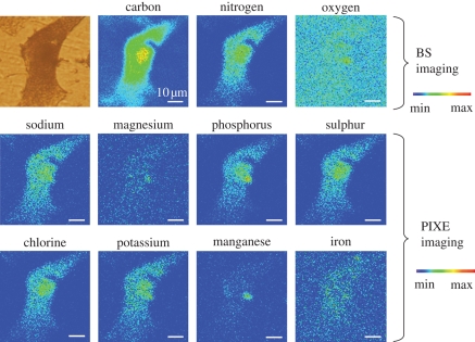
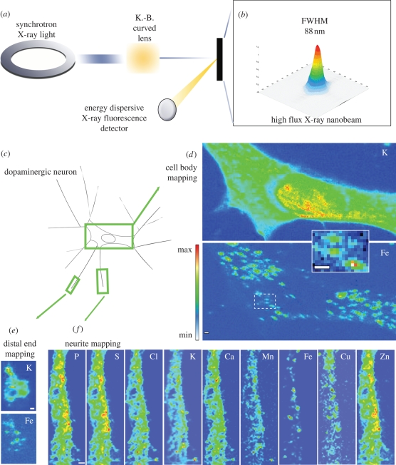
The direct detection of biologically relevant metals in single cells and of their speciation is a challenging task that requires sophisticated analytical developments. The aim of this article is to present the recent achievements in the field of cellular chemical element imaging, and direct speciation analysis, using proton and synchrotron radiation X-ray micro- and nano-analysis. The recent improvements in focusing optics for MeV-accelerated particles and keV X-rays allow application to chemical element analysis in subcellular compartments. The imaging and quantification of trace elements in single cells can be obtained using particle-induced X-ray emission (PIXE). The combination of PIXE with backscattering spectrometry and scanning transmission ion microscopy provides a high accuracy in elemental quantification of cellular organelles. On the other hand, synchrotron radiation X-ray fluorescence provides chemical element imaging with less than 100 nm spatial resolution. Moreover, synchrotron radiation offers the unique capability of spatially resolved chemical speciation using micro-X-ray absorption spectroscopy. The potential of these methods in biomedical investigations will be illustrated with examples of application in the fields of cellular toxicology, and pharmacology, bio-metals and metal-based nano-particles.


Figures

Figure 1.
Figure 1.
Multi-element imaging of a single PC12 cell exposed to MnCl2 obtained at the AIFIRA facility. Carbon, nitrogen and oxygen maps were imaged using micro-BS analysis, whereas trace elements of higher Z were imaged using micro-PIXE analysis. The chemical element maps show that Mn accumulates within the perinuclear region of the cell and is colocalized with Mg. Scale bar, 10 µm.
Figure 2.
Figure 2.
(a) The synchrotron XRF nanoprobe end-station installed at ESRF, on the ID22 beamline, was designed to provide a high flux (1012 ph s−1) of hard X-rays (i.e. 17 keV) with a beam size of less than 90 nm (FWHM, full width at half maximum) using a Kirkpatrick–Baez-type curved lens. The intensity distribution in the focal plane is shown in (b); dopamine-producing cells were exposed in vitro to FeSO4 for 24 h (c). Chemical element distributions were recorded on distinct cellular areas such as cell bodies (d), distal ends (e) and neurite outgrowths (f). Min–max range bar units are arbitrary. All scale bars, 1 µm. Iron was found in 200–300 nm structures in the cytosol, neurite outgrowths and distal ends, but not in the nucleus (Ortega et al. 2007).
Figure 3.
Figure 3.
Normalized XANES spectra at arsenic K-absorption edge in the cytosol, mitochondrial network, nucleus and lysosomes of HepG2 cells exposed to As(OH)3. Light microscopy of a single cell (a); epifluorescence microscopy of the same cell showing in green the mitochondrial network (labelled with rhodamine 123) and in blue the lysosomes (labelled with lysotracker) (b); micro-XRF distribution of potassium (c) and arsenic (d). XANES spectra in the subcellular compartments show that the main arsenic oxidation state is As(III) in all organelles, with a mixture of trivalent and pentavalent arsenic in the nucleus (e).

References

    1. Bacquart T., Devès G., Carmona A., Tucoulou R., Bohic S., Ortega R. 2007. Subcellular speciation analysis of trace element oxidation states using synchrotron radiation micro-X-ray absorption near edge structure. Anal. Chem. 79, 7353–7359. (10.1021/ac0711135) - DOI - PubMed
    1. Barberet Ph., Incerti S., Andersson F., Delalee F., Serani L., Moretto Ph. 2009. Technical description of the CENBG nanobeam line. Nucl. Instr. Methods B 267, 2003–2007. (10.1016/j.nimb.2009.03.077) - DOI
    1. Bench G., Grant P. G., Ueda D. L., Autry-Conwell S. A., Hou Y., Boggan J. E. 2003. Assessment of proton microbeam analysis of 11B for quantitative microdistribution analysis of boronated neutron capture agents in biological tissues. Radiat. Res. 160, 667–676. (10.1667/RR3085) - DOI - PubMed
    1. Bohic S., Murphy K., Paulus W., Cloetens P., Salomé M., Susini J., Double K. 2008. Intracellular chemical imaging of the developmental phases of human neuromelanin using synchrotron X-ray microspectroscopy. Anal. Chem. 80, 9557–9566. (10.1021/ac801817k) - DOI - PubMed
    1. Bussy C., et al. 2008. Carbon nanotubes in macrophages: imaging and chemical analysis by X-ray fluorescence microscopy. Nano Lett. 8, 2659–2663. (10.1021/nl800914m) - DOI - PubMed

LinkOut - more resources