. 2009 Oct;83(19):10058-74.
doi: 10.1128/JVI.02418-08. Epub 2009 Jul 15.

Class III phosphatidylinositol 4-kinase alpha and beta are novel host factor regulators of hepatitis C virus replication

Affiliations

Class III phosphatidylinositol 4-kinase alpha and beta are novel host factor regulators of hepatitis C virus replication

Jason Borawski et al. J Virol. 2009 Oct.

Abstract

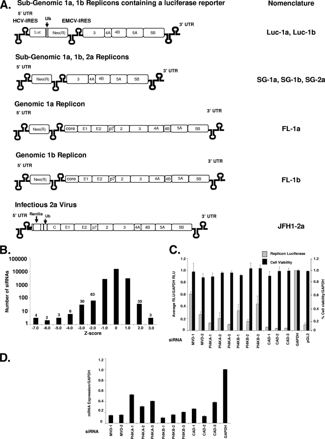
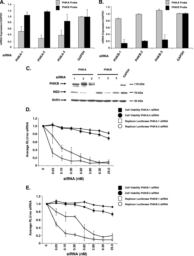
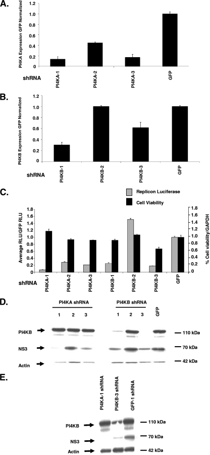
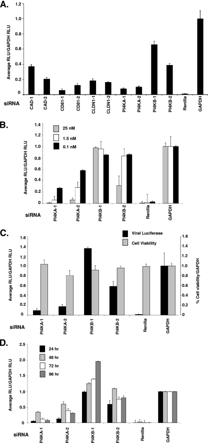
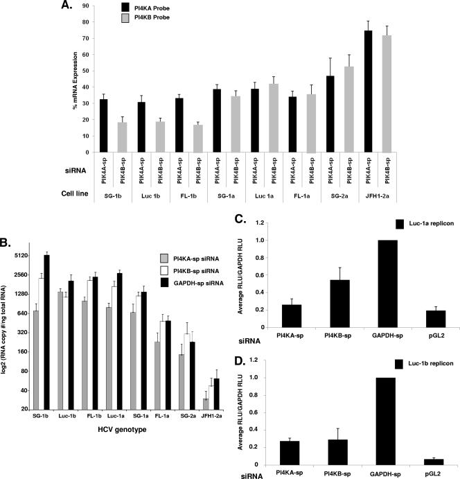
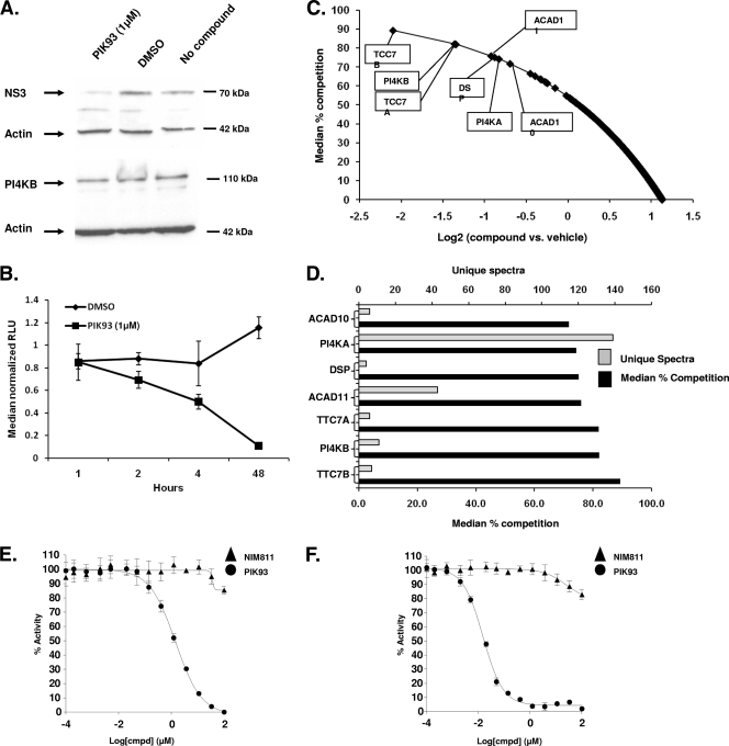
Host factor pathways are known to be essential for hepatitis C virus (HCV) infection and replication in human liver cells. To search for novel host factor proteins required for HCV replication, we screened a subgenomic genotype 1b replicon cell line (Luc-1b) with a kinome and druggable collection of 20,779 siRNAs. We identified and validated several enzymes required for HCV replication, including class III phosphatidylinositol 4-kinases (PI4KA and PI4KB), carbamoyl-phosphate synthetase 2, aspartate transcarbamylase, and dihydroorotase (CAD), and mevalonate (diphospho) decarboxylase. Knockdown of PI4KA could inhibit the replication and/or HCV RNA levels of the two subgenomic genotype 1b clones (SG-1b and Luc-1b), two subgenomic genotype 1a clones (SG-1a and Luc-1a), JFH-1 genotype 2a infectious virus (JFH1-2a), and the genomic genotype 1a (FL-1a) replicon. In contrast, PI4KB knockdown inhibited replication and/or HCV RNA levels of Luc-1b, SG-1b, and Luc-1a replicons. The small molecule inhibitor, PIK93, was found to block subgenomic genotype 1b (Luc-1b), subgenomic genotype 1a (Luc-1a), and genomic genotype 2a (JFH1-2a) infectious virus replication in the nanomolar range. PIK93 was characterized by using quantitative chemical proteomics and in vitro biochemical assays to demonstrate PIK93 is a bone fide PI4KA and PI4KB inhibitor. Our data demonstrate that genetic or pharmacological modulation of PI4KA and PI4KB inhibits multiple genotypes of HCV and represents a novel druggable class of therapeutic targets for HCV infection.


Figures

FIG. 1.
FIG. 1.
Genome-scale siRNA screen in HCV replicon cells. (A) Model of the HCV replicons used in the present study: the subgenomic genotype 1a luciferase replicon (Luc-1a) and the subgenomic genotype 1b luciferase replicon (Luc-1b), both of which contain a Firefly luciferase gene; the subgenomic genotype 1a replicon (SG-1a), the subgenomic genotype 1b replicon (SG-1b), the subgenomic genotype 2a replicon (SG-2a), the genomic genotype 1a replicon (FL-1a), and the genomic genotype 1b replicon (FL-1b), all of which contain a neomycin selectable marker; and the genomic genotype 2a infectious virus (JFH1-2a), which contains a Renilla luciferase gene. (B) The Huh7 stable HCV replicon cells were screened by high-throughput lipid-mediated transfection using a SMARTpool kinome (779 genes) and a druggable genome (four siRNAs targeting 5,000 genes) at 25 nM siRNA per well. The distribution of siRNAs is presented as a Z-score histogram. The frequency of hits at each Z-score is presented above each column. Including the luciferase siRNA, 108 siRNAs scored below a Z-score of −2, whereas 38 siRNAs scored above 2.0. (C) The top-scoring siRNA inhibitors were validated by using multiple independent siRNAs (see Table S3 in the supplemental material for sequences) after a 72-h transfection into the Luc-1b cells. Both luciferase (░⃞) and cell viability (▪) were determined and normalized to the negative control GAPDH siRNA. (D) mRNA levels were quantified in the siRNA-treated cells (in parallel with panel C) using RT-PCR, based on the internal control actin mRNA. The data were normalized to GAPDH siRNA-treated cells to control for transfection effects on total cellular mRNA levels.
FIG. 2.
FIG. 2.
Validation of PI4KA and PI4KB siRNAs. (A and B) Each PI4K siRNA was analyzed by RT-PCR to determine whether the siRNAs were isoform specific. After siRNA transfection with PI4KA (A) and PI4KB (B) siRNAs, the mRNA levels were measured using both a PI4KA and a PI4KB TaqMan probe. (C) PI4KB and NS3 protein levels were measured in replicon cell lysates after a 72-h siRNA transfection with 25 nM siRNA. (D and E) Luc-1b cells were transfected with a dose response of PI4KA-1 (□) and PI4KA-2 (○) (D) or PI4KB-1 (□) and PI4KB-2 (○) (E) siRNAs, luciferase levels (open symbols) and cell viability (solid symbols) were quantified and normalized to the siRNA-free treatment (the zero concentration). RT-PCR was also confirmed across the dose response in Fig. S3A and B in the supplemental material. The RT-PCR data are presented as normalized to GAPDH and are representative of three independent experiments, along with the standard deviations.
FIG. 3.
FIG. 3.
Stable silencing of PI4KA and PI4KB in Luc-1b replicon cells. (A and B) Three shRNAs targeting PI4KA or PI4KB were stably transduced into Luc-1b cells (on puromycin for 2 weeks), and the mRNA levels were quantified by using RT-PCR for PI4KA (A) and PI4KB (B). (C) In parallel, the PI4KA or PI4KB stably transduced cells were analyzed for luciferase signal 96 h after plating (░⃞), and ATP levels were quantified as a surrogate for cell viability (▪). The PI4KB and NS3 protein levels were measured in stable replicon lysates after 96 h (D) or 3 weeks (E) on puromycin selection. All data are presented as normalized to GFP shRNAs and are representative of three independent experiments. The standard deviations are indicated.
FIG. 4.
FIG. 4.
PI4KA and PI4KB siRNAs inhibit JFH-1 infectious virus. (A) A set of SMARTpool siRNAs known to affect HCV entry were transfected into JFH1-2a-infected cells at 25 nM, and the Renilla expression was quantified at 72 h. (B) siRNAs were transfected over a dose response (25, 1.5, and 0.1 nM, as indicated) into Huh7.5 cells for 24 h prior to infection with the JFH-1 virus. After 48 h, the cells were harvested for Renilla luciferase quantification. The mRNA levels were analyzed in the Huh7.5 cells at 72 h after a 25 nM siRNA transfection and viral infection (data not shown). (C) Huh7.5 cells were first infected with JFH-1 for 24 h prior to siRNA transfection for 72 h, and the levels of viral luciferase (▪) and cell viability (░⃞) were measured. (D) Conditioned supernatants from the JFH1-2a-infected Huh 7.5 cells in panel C were collected and added back onto naive Huh7.5 cells. Luciferase was measured from 24 to 96 h (24, 48, 72, and 96 h, as indicated) after supernatant addition to measure reinfection rates. All data are presented as normalized to GAPDH siRNAs and are representative of three independent experiments. The standard deviations are indicated.
FIG. 5.
FIG. 5.
QRT-PCR analysis of PI4KA-sp and PI4KB-sp siRNAs across multiple HCV replicons. Eight HCV replicons (SG-1b, Luc-1b, FL-1b, Luc-1a, SG-1a, FL-1a, SG-2a, and JFH1-2a) were transfected with 25 nM concentrations of PI4KA-sp, PI4KB-sp, and GAPDH-sp siRNAs for 72 h, and mRNA was isolated. (A) mRNA knockdown of PI4KA and PI4KB was determined in each transfection by RT-PCR and is shown as the percent mRNA expression for each cell line. (B) The HCV RNA copy number per ng of total RNA was determined as previously described (37). The HCV copy number data was log2 transformed to compare all of the replicons in parallel. The copy number was the average of three independent transfections. The standard deviations are indicated. (C and D) The PI4KA-sp, PI4KB-sp, and GAPDH-sp siRNA-treated (25 nM) Luc-1a cells (C) and Luc-1b cells (D) were analyzed for luciferase levels after a 72-h transfection.
FIG. 6.
FIG. 6.
Biochemistry of PIK93 in Luc-1b cells. (A) PI4KB, NS3, and β-actin protein levels were analyzed in the Luc-1b cells by Western blot after a 1 μM 48-h incubation with PIK93, DMSO, or without compound. (B) The luciferase activity was also determined in parallel the Luc-1b cells (panel A) after treatment with 1 μM PIK93 over a 48-h time course. (C) Affinity chromatography in-lysate competition experiments were performed in the HCV replicon cells with 10 μM PIK93 and DMSO (affinity matrix supplied in Fig. S4 in the supplemental material). Peptides that competed PIK93 greater than 70% are highlighted in the boxes. All peptides competed from the column are indicated in Table S4 in the supplemental material. (D) Differentially competed proteins are shown as the percent competition verses the DMSO control lysates (▪) and the unique spectra for each protein (░⃞). (E and F) PI4KA (E) or PI4KB (F) protein was preincubated for 10 min at 14 different concentrations of PIK93 (•) or NIM811 (▴) before the addition of ATP and phosphatidylinositol. Depletion of NADH fluorescence was measured for 5 min, and the percent activity was calculated from the initial rates of the inhibited reactions relative to the uninhibited control.

References

    1. Ahn, J., K. Chung, D. Kim, M. Won, L. Kim, K. Kim, M. Nam, S. Choi, H. Kim, M. Yoon, S. Chae, and K. Hoe. 2004. Systematic identification of hepatocellular proteins interacting with NS5A of the hepatitis C virus. J. Biochem. Mol. Biol. 37:741-748. - PubMed
    1. Aizaki, H., K. Lee, V. Sung, H. Ishiko, and M. Lai. 2004. Characterization of the hepatitis C virus RNA replication complex associated with lipid rafts. Virology 324:450-461. - PubMed
    1. Bader, T., J. Fazili, M. Madhoun, C. Aston, D. Hughes, S. Rizvi, K. Seres, and M. Hasan. 2008. Fluvastatin inhibits hepatitis C replication in humans. Am. J. Gastroenterol. 103:1383-1389. - PubMed
    1. Balla, A., Y. Kim, P. Varnai, Z. Szentpetery, Z. Knight, K. Shokat, and T. Balla. 2008. Maintenance of hormone-sensitive phosphoinositide pools in the plasma membrane requires phosphatidylinositol 4-kinase IIIα. Mol. Biol. Cell 19:711-721. - PMC - PubMed
    1. Balla, A., G. Tuymetova, A. Tsiomenko, P. Várnai, and T. Balla. 2005. A plasma membrane pool of phosphatidylinositol 4-phosphate is generated by phosphatidylinositol 4-kinase type-III alpha: studies with the PH domains of the oxysterol binding protein and FAPP1. Mol. Biol. Cell 16:1282-1295. - PMC - PubMed

LinkOut - more resources