. 2009 Oct;83(19):9923-39.
doi: 10.1128/JVI.00066-09. Epub 2009 Jul 15.

Characterization of hepatitis C virus core protein multimerization and membrane envelopment: revelation of a cascade of core-membrane interactions

Affiliations

Characterization of hepatitis C virus core protein multimerization and membrane envelopment: revelation of a cascade of core-membrane interactions

Li-Shuang Ai et al. J Virol. 2009 Oct.

Abstract

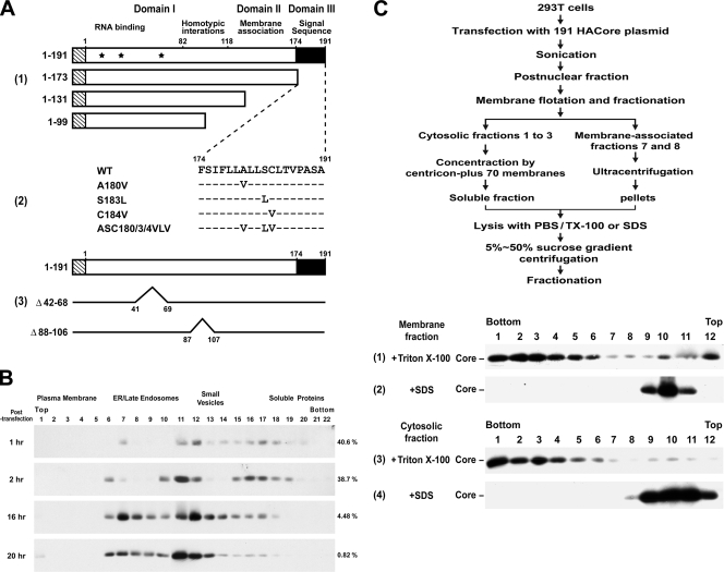
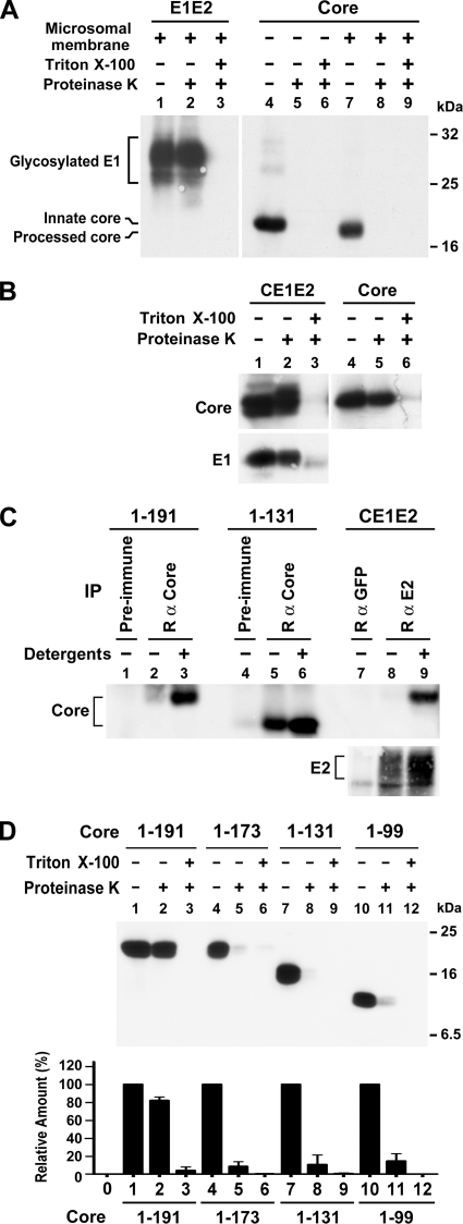
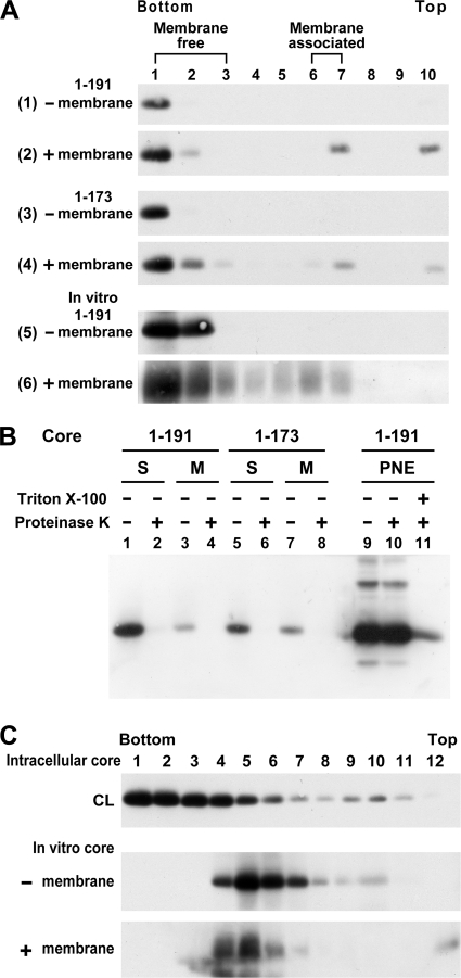
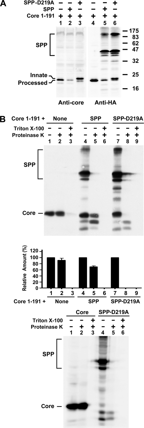
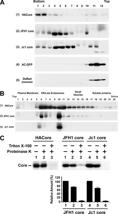
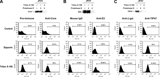
The molecular basis underlying hepatitis C virus (HCV) core protein maturation and morphogenesis remains elusive. We characterized the concerted events associated with core protein multimerization and interaction with membranes. Analyses of core proteins expressed from a subgenomic system showed that the signal sequence located between the core and envelope glycoprotein E1 is critical for core association with endoplasmic reticula (ER)/late endosomes and the core's envelopment by membranes, which was judged by the core's acquisition of resistance to proteinase K digestion. Despite exerting an inhibitory effect on the core's association with membranes, (Z-LL)(2)-ketone, a specific inhibitor of signal peptide peptidase (SPP), did not affect core multimeric complex formation, suggesting that oligomeric core complex formation proceeds prior to or upon core attachment to membranes. Protease-resistant core complexes that contained both innate and processed proteins were detected in the presence of (Z-LL)(2)-ketone, implying that core envelopment occurs after intramembrane cleavage. Mutations of the core that prevent signal peptide cleavage or coexpression with an SPP loss-of-function D219A mutant decreased the core's envelopment, demonstrating that SPP-mediated cleavage is required for core envelopment. Analyses of core mutants with a deletion in domain I revealed that this domain contains sequences crucial for core envelopment. The core proteins expressed by infectious JFH1 and Jc1 RNAs in Huh7 cells also assembled into a multimeric complex, associated with ER/late-endosomal membranes, and were enveloped by membranes. Treatment with (Z-LL)(2)-ketone or coexpression with D219A mutant SPP interfered with both core envelopment and infectious HCV production, indicating a critical role of core envelopment in HCV morphogenesis. The results provide mechanistic insights into the sequential and coordinated processes during the association of the HCV core protein with membranes in the early phase of virus maturation and morphogenesis.


Figures

FIG. 1.
FIG. 1.
Construction of core plasmids and analyses of membrane-bound and -free core complexes. (A) pCAGGS-based plasmids encoding residues 1 to 191 of the genotype 1a H77 strain core protein and C-terminal-truncated mutants tagged with the influenza virus HA epitope at the N terminus, as shown by the striped rectangles, were constructed as described in Materials and Methods. Domain I, which contains three nuclear localization sequences (located at residues 6 to 13, 38 to 43, and 59 to 71, respectively, and marked with asterisks) and a region involved in core homotypic interaction; domain II, which contains hydrophobic sequences for membrane association; and domain III, which serves as the signal sequence for E1, are also indicated (panel 1). pCAGGS-HACore-based mutant plasmids encoding substitutions in the signal peptide at the residues as indicated were also constructed (panel 2). Dashes indicate that the residue in that position of the mutant core protein is identical to that of the WT core. The Δ88-106 and Δ42-68 mutants, and their corresponding deleted regions are shown in panel 3. (B) 293T cells were transfected with the 191 core-expressing plasmid, and the calcium phosphate-DNA complexes were washed away 4 h posttransfection. Cells were then incubated at 37°C for different times as indicated and harvested, and the postnuclear fractions were subjected to Optiprep subcellular fractionation, followed by Western blot analysis with an HA MAb. The blots were scanned, and the percentages of core proteins distributed into soluble fractions 16 to 22 of the total core populations are indicated on the right of each panel. (C) The steady-state 191 core proteins localized in membrane-associated fractions 7 and 8 after the membrane flotation assay (data not shown) were combined, mixed with an equal volume of cold TE buffer, and then centrifuged in a Beckman SW41 rotor at 36,000 rpm for 2 h. The pelleted membranes were lysed with 0.5 ml of PBS containing 1% Triton X-100 (panel 1) or SDS (panel 2) at 37°C for 10 min prior to 5 to 50% sucrose gradient centrifugation. The combined cytosolic fractions 1 to 3 after the membrane flotation assay (data not shown) were diluted with 6 volumes of cold TE buffer and concentrated by Amicon Centricon plus-70 membranes (Millipore, Bedford, MA). The samples (0.5 ml) were treated with a final concentration of 1% Triton X-100 (panel 3) or SDS (panel 4) before 5 to 50% sucrose gradient centrifugation. The scheme illustrating these procedures is shown at the top.
FIG. 2.
FIG. 2.
Assessment of core envelopment by membrane protection and immunoprecipitation assays. (A) E1E2 and core proteins translated in vitro from pcDNA3-E1E2 and pcDNA3-HACore, respectively, in the presence or absence of canine pancreatic microsomal membranes were subjected to a membrane protection assay. Proteins were resolved by SDS-PAGE, followed by Western blotting with E1- and HA-specific MAbs, respectively. (B) Equal volumes of postnuclear fractions obtained from 293T cells transfected with pCAGGS-HACE1E2 or pCAGGS-HACore were subjected to a membrane protection assay, and samples were analyzed by Western blotting with HA- and E1-specific MAbs, respectively. (C) Postnuclear fractions obtained from 293T cells expressing core or CE1E2 proteins were left untreated or treated with 1% each of NP-40 and sodium deoxycholate prior to immunoprecipitation with the core or E2-specific antibodies as indicated. Incubation with appropriate rabbit preimmune serum or purified rabbit anti-GFP was used as the control. The precipitated antigens were analyzed by Western blotting with MAbs directed against HA and E2, respectively. (D) Postnuclear samples from cells transfected with each core plasmid were analyzed by a membrane protection assay followed by Western blotting with the HA MAb (top panel). The density of bands corresponding to the HA core proteins in each treatment was scanned and quantified. The percentages of core levels detected in samples treated with proteinases K alone or with detergent and protease relative to that of an untreated sample were determined. The diagram represents results from three independent studies with the standard deviation shown (bottom panel).
FIG. 3.
FIG. 3.
In vitro membrane-binding ability and multimerization of core complexes. (A) 293T cells expressing the 1-191 (panels 1 and 2) and 1-173 (panels 3 and 4) core proteins were lysed with 1% each of NP-40 and sodium deoxycholate and then fractionated by 5 to 50% sucrose gradient centrifugation. The bottom fractions containing the high-ordered, multimeric core complexes were diluted with PBS and ultracentrifuged. After resuspension with PBS, the core complexes were divided into two portions: one left untreated (panels 1 and 3, respectively) and the other incubated with canine pancreatic microsomal membrane (panels 2 and 4, respectively). The mixtures were then analyzed by a membrane flotation assay, followed by Western blotting with the HA MAb. The 191 core protein translated in vitro from pcDNA3-HACore in the absence (panel 5) or presence (panel 6) of microsomal membranes was also resolved by membrane flotation. (B) The pooled membrane-free fractions 1 to 3 and membrane-associated fractions 7 and 8, marked as S and M, respectively, obtained from panels 2 and 4 in panel A were concentrated and treated with or without proteinase K prior to Western blotting (lanes 1 to 8). Postnuclear fractions containing the processed 191 core were simultaneously digested with proteinase K in the presence or absence of detergent (lanes 9 to 11) and were used as the controls. (C) Detergent-treated cell lysates (marked as CL) containing the 191 core and core synthesized in vitro in the absence or presence of microsomal membranes were analyzed by 5 to 50% sucrose gradient centrifugation.
FIG. 4.
FIG. 4.
Characterization of the 191 core synthesized in the presence of (Z-LL)2-ketone. (A) 293T cells expressing the 191 core were treated with or without 100 μM (Z-LL)2-ketone at 37°C for 14 h, and the postnuclear extracts were analyzed by Optiprep-based subcellular fractionation. Migration of the innate p23 and processed p21 core proteins is marked. Percentages of core proteins distributed in soluble fractions 16 to 22 of the total core populations are indicated on the right of each panel. (B) 293T cells expressing the 191 core were treated with or without 20 μM (Z-LL)2-ketone at 37°C for 20 h, and the postnuclear extracts were subjected to a proteinase K digestion assay. The amount of the core synthesized in the presence of the SPP inhibitor was adjusted for better comparison of the protease resistance to that of the core expressed without the SPP inhibitor. (C) Cells expressing the 191 core were treated with 20 μM (Z-LL)2-ketone for 20 h or untreated, and cell lysates were subjected to 5 to 50% sucrose gradient centrifugation.
FIG. 5.
FIG. 5.
Analyses of core proteins with mutations in the signal sequence. (A) Postnuclear fractions of cells expressing the 191 WT core or core mutants were subjected to Optiprep-based subcellular fractionation. Percentages of core proteins partitioned into soluble fractions 16 to 22 of the total core populations are indicated on the right of each panel. (B) Postnuclear fractions obtained from cells expressing various core proteins were assessed by a membrane protection assay (top panel). Blots were scanned for the intensity of the core band. In each case, the relative amounts of the core treated with proteinase K or with detergent and protease relative to that of the core left untreated were assessed. The diagram represents data from three separate experiments with the standard deviation shown (bottom panel).
FIG. 6.
FIG. 6.
Analyses of the 191 core coexpressed with the WT or D219A mutant SPP. (A) 293T cells were cotransfected with the 191 core plasmid in the presence of pcDNA3, the WT, or D219A mutant SPP plasmid. Cell lysates were analyzed by Western blotting with the rabbit anti-core and HA MAb, respectively. (B) Postnuclear fractions from cells expressing the core alone or coexpressing the core and WT or mutant SPP were subjected to a membrane protection assay, followed by Western blotting with the HA MAb (top panel). The results from three individual studies were quantified, and the average values with the standard deviation are also shown (middle panel). Postnuclear fractions from cells transfected with the 191 core or D219A SPP mutant plasmid were analyzed by a proteinase K digestion assay (bottom panel).
FIG. 7.
FIG. 7.
Characterization of core mutants with deletions in domain I. (A) Detergent-treated lysates from 293T cells expressing each of the 191 and deletion mutants were subjected to 5 to 50% sucrose linear density gradient centrifugation followed by Western blotting with the HA MAb. (B) Postnuclear fractions obtained from cells expressing each of indicated core proteins were analyzed by Optiprep subcellular fractionation. (C) 293T cells were cotransfected with the core plasmids as indicated in the presence of pcDNA3 or D219A mutant SPP plasmid. Cell lysates were analyzed by Western blotting with the HA MAb. (D) Postnuclear fractions from cells expressing each of the core proteins were subjected to a membrane protection assay, and a representative immunoblot is shown (top panel). The results from three separate analyses were quantified, and the average values with the standard deviation are shown (bottom panel).
FIG. 8.
FIG. 8.
Characterization of core proteins expressed from infectious HCV RNAs. (A) Huh7 cells were transfected with the 191 pCAGGS-HACore plasmid, JFH1 and Jc1 RNAs, pAcGFP-N1, and pDsRed-monomeric-C1 (Clontech Laboratories, Mountain View, CA), respectively. Transfected cells were lysed with 1% each of NP-40 and sodium deoxycholate, and cell lysates were then subjected to 5 to 50% sucrose density gradient centrifugation, followed by Western blotting with the core MAb (panels 1 to 3), rabbit anti-GFP (panel 4), and MAb for DsRed (panel 5), respectively. (B) Huh7 cells were transfected with the 191 core plasmid, and JFH1 and Jc1 RNAs, respectively, and postnuclear fractions were analyzed by Optiprep-based subcellular fractionation followed by Western blotting with the core MAb. (C) Postnuclear fractions obtained from Huh7 cells transfected with the 191 core plasmid, and JFH1 and Jc1 RNAs, respectively, were analyzed by a membrane protection assay (top panel). The percentages of resistance to proteinase K digestion in the presence or absence of detergent for the JFH1 and Jc1 core proteins were quantified, and the results from four independent studies with the standard deviation are shown (bottom panel).
FIG. 9.
FIG. 9.
Assessment of core envelopment by differential membrane permeabilization. (A) Huh7 cells transfected with JFH1 RNA were incubated with PBS, or permeabilized by saponin or Triton X-100. After blocking, cells from each treatment were separately incubated with 5 μl each of rabbit preimmune or anti-core serum, which was followed by incubation with FITC-conjugated goat anti-rabbit IgG. The immunostained cells were quantified by flow cytometry. The specific levels of anti-core-positive cells revealed by saponin and Triton X-100, designated as S and T values, respectively, were obtained by subtracting the background level with preimmune antibody incubation from those with anti-core incubation. The percentage of core envelopment is defined as [(T − S)]/T × 100%. Two separate analyses were performed with similar results. The data from a representative set are thus shown. The postnuclear fraction from the same experiment was also subjected to the membrane protection assay. (B and C) JFH1 RNA-transfected Huh7 cells were divided into two parts for the studies as shown. In each case, a portion of cells was prepared for postnuclear fractions which were then subjected to the HCV E2 protein (B) and TIP47 (C) membrane protection assays, respectively. Another portion of cells was further divided into three parts for the differential membrane permeabilization assay as described in panel A. Cells from each treatment were then separately incubated with 1 μg each of affinity-purified control mouse IgG or an E2 MAb (B) or with 1 μg each of goat anti-β-Gal or goat anti-TIP47 IgG (C) followed by incubation with FITC-linked rabbit anti-mouse (B) or rabbit anti-goat IgGs (C). The immunostained cells were then analyzed by FACS. In panels B and C, three independent analyses were performed with similar results. Thus, data from a representative set are shown.
FIG. 10.
FIG. 10.
Analysis of the effects of (Z-LL)2-ketone and D219A mutant SPP on core envelopment and infectious virus production. (A) Huh7 cells were first transfected with 10 μg of JFH1 RNA, and at 16 h posttransfection the cell cultures were supplemented with or without 20 μM (Z-LL)2-ketone. At 64 h after the beginning of transfection, the cells were harvested, and postnuclear fractions were subjected to a proteinase K digestion assay. The diagram represents results from three independent studies with the standard deviation shown (top and middle panels). The viral infectivity of cell-free supernatants obtained was determined as focus-forming units (FFU) per milliliter, and the results from three independent studies with the standard deviation are shown (bottom panel). (B) JFH1 RNA-transfected Huh7 cells were treated with or without (Z-LL)2-ketone, and transfected cells were then analyzed by differential membrane permeabilization, followed by FACS. The data from a representative set are shown. The degree of core envelopment in the presence or absence of the SPP inhibitor was calculated as described in Fig. 9A. The diagram represents results from three independent studies with the standard deviation shown. (C) Huh7 cells were first transfected with 10 μg of JFH1 RNA and 16 h posttransfection cells were separately transfected with 10 μg of the vector, marked as “Mock”, or the D219A mutant SPP plasmid. At 64 h after the beginning of transfection, postnuclear fractions were prepared and subjected to a proteinase K digestion assay (top and middle panel), and culture supernatants were titrated for viral infectivity (bottom panel). In each case, the diagram represents results from three independent studies with the standard deviation shown. Differences between samples treated with (Z-LL)2-ketone or cotransfected with D219A plasmid and controls were determined by using the Student t test: P < 0.05 (A and C) and P = 0.0001 (B).
FIG. 11.
FIG. 11.
Model for the concerted events of core interaction with and envelopment by ER membranes. The 1-191 core is synthesized in the cytoplasm and assembled into an oligomeric structure before or upon attachment to the cytoplasmic side of the ER membrane via the core C-terminal signal peptide. The core oligomers may continue to multimerize into a high-ordered, structural complex on the cytoplasmic surface of the ER membrane. The signal peptide is then cleaved off by intramembrane SPP, which is subsequently followed by envelopment of core complex into the lumen of ER membranes. The SPP inhibitor, (Z-LL)2-ketone, interferes with both signal sequence processing and attachment of the core complex to the ER membrane.

Similar articles

Cited by

References

    1. Acosta-Rivero, N., A. Musacchio, L. Lorenzo, C. Alvarez, and J. Morales. 2002. Processing of the hepatitis C virus precursor protein expressed in the methylotrophic yeast Pichia pastoris. Biochem. Biophys. Res. Commun. 295:81-84. - PubMed
    1. Acosta-Rivero, N., A. Rodriguez, A. Musacchio, V. Falcon, V. M. Suarez, G. Martinez, I. Guerra, D. Paz-Lago, Y. Morera, M. C. de la Rosa, J. Morales-Grillo, and S. Duenas-Carrera. 2004. In vitro assembly into virus-like particles is an intrinsic quality of Pichia pastoris derived HCV core protein. Biochem. Biophys. Res. Commun. 325:68-74. - PubMed
    1. Barba, G., F. Harper, T. Harada, M. Kohara, S. Goulinet, Y. Matsuura, G. Eder, Z. Schaff, M. J. Chapman, T. Miyamura, and C. Brechot. 1997. Hepatitis C virus core protein shows a cytoplasmic localization and associates to cellular lipid storage droplets. Proc. Natl. Acad. Sci. USA 94:1200-1205. - PMC - PubMed
    1. Bartenschlager, R., M. Frese, and T. Pietschmann. 2004. Novel insights into hepatitis C virus replication and persistence. Adv. Virus Res. 63:71-180. - PubMed
    1. Bartenschlager, R., and V. Lohmann. 2000. Replication of hepatitis C virus. J. Gen. Virol. 81:1631-1648. - PubMed

Publication types

LinkOut - more resources