. 2009 Aug;136(16):2815-23.
doi: 10.1242/dev.038620. Epub 2009 Jul 15.

Sox2-positive dermal papilla cells specify hair follicle type in mammalian epidermis

Affiliations

Sox2-positive dermal papilla cells specify hair follicle type in mammalian epidermis

Ryan R Driskell et al. Development. 2009 Aug.

Abstract

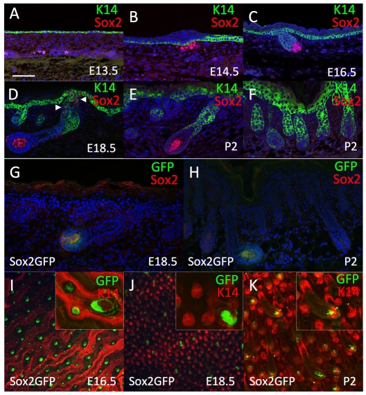
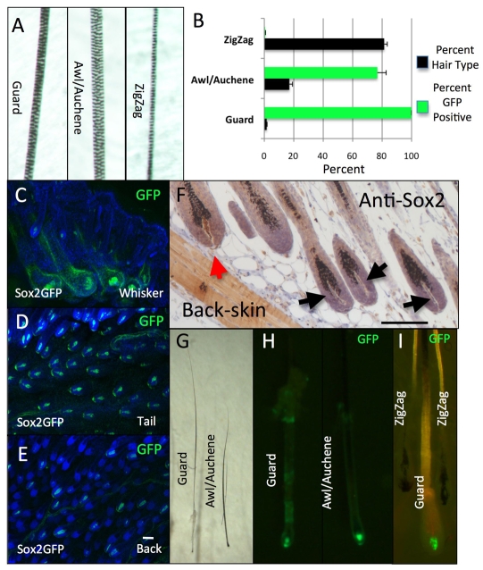
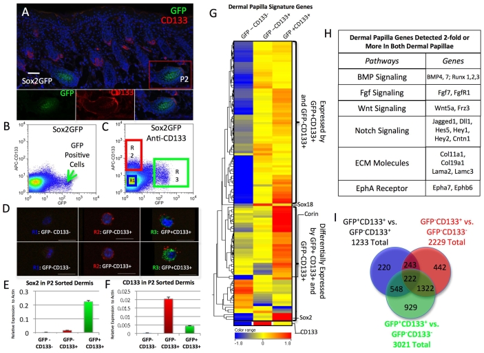
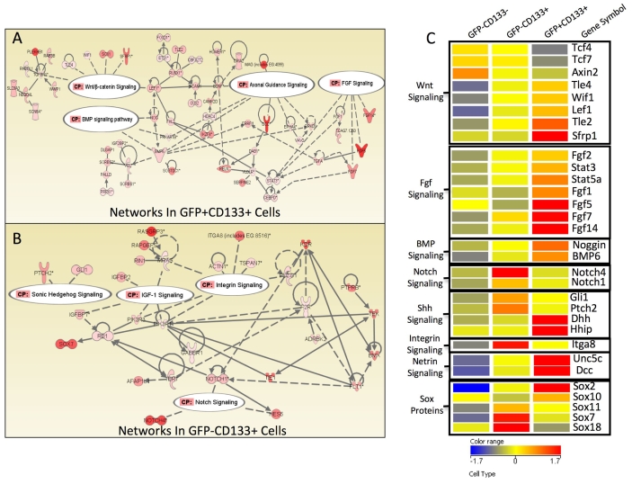
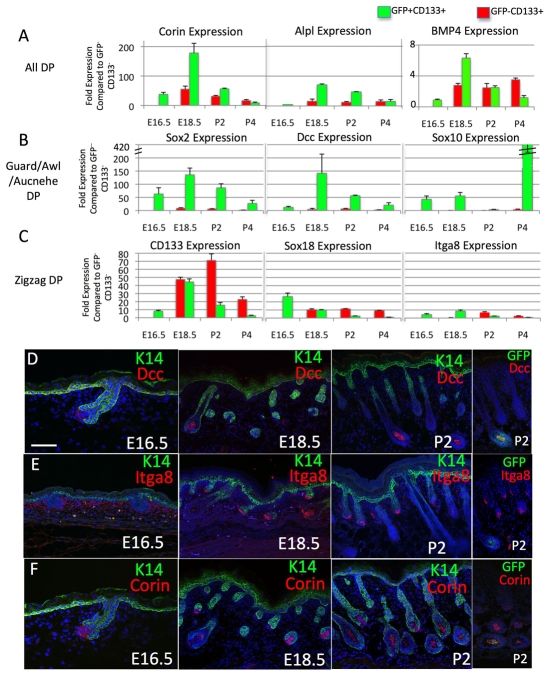
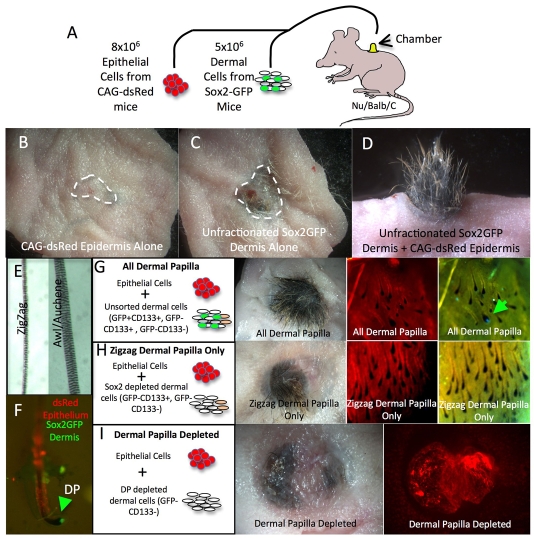
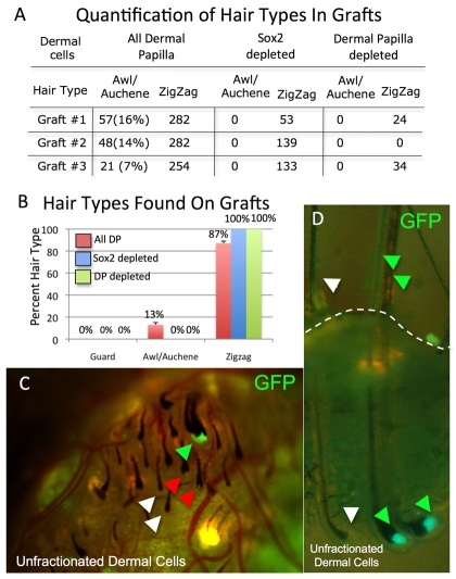
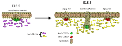
The dermal papilla comprises the specialised mesenchymal cells at the base of the hair follicle. Communication between dermal papilla cells and the overlying epithelium is essential for differentiation of the hair follicle lineages. We report that Sox2 is expressed in all dermal papillae at E16.5, but from E18.5 onwards expression is confined to a subset of dermal papillae. In postnatal skin, Sox2 is only expressed in the dermal papillae of guard/awl/auchene follicles, whereas CD133 is expressed both in guard/awl/auchene and in zigzag dermal papillae. Using transgenic mice that express GFP under the control of the Sox2 promoter, we isolated Sox2(+) (GFP(+)) CD133(+) cells and compared them with Sox2(-) (GFP(-)) CD133(+) dermal papilla cells. In addition to the 'core' dermal papilla gene signature, each subpopulation expressed distinct sets of genes. GFP(+) CD133(+) cells had upregulated Wnt, FGF and BMP pathways and expressed neural crest markers. In GFP(-) CD133(+) cells, the hedgehog, IGF, Notch and integrin pathways were prominent. In skin reconstitution assays, hair follicles failed to form when dermis was depleted of both GFP(+) CD133(+) and GFP(-) CD133(+) cells. In the absence of GFP(+) CD133(+) cells, awl/auchene hairs failed to form and only zigzag hairs were found. We have thus demonstrated a previously unrecognised heterogeneity in dermal papilla cells and shown that Sox2-positive cells specify particular hair follicle types.


Figures

Fig. 1.
Fig. 1.
Sox2 expression is localised to the dermal papilla of specific hair follicles. (A-F) Paraffin sections of E13.5 (A), E14.5 (B), E16.5 (C), E18.5 (D) and P2 (E,F) wild-type skin immunolabelled with antibodies to Sox2 (red) and keratin 14 (green), with DAPI nuclear counterstain (blue). Arrows in D indicate Sox2-positive Merkel cells. (G,H) Cryosections of Sox2GFP skin immunolabelled for Sox2 and GFP at E18.5 (G) and P2 (H), with DAPI nuclear counterstain (blue). (I-K) Confocal micrographs of Sox2GFP transgenic mouse skin whole mounts at E16.5 (I), E18.5 (J) and P2 (K) immunolabelled with anti-keratin 14 (red). Inserts show higher magnification views of dermal papillae. Scale bar: 100 μm.
Fig. 2.
Fig. 2.
Sox2 expression is restricted to guard/awl/auchene dermal papillae. (A) Bright-field images showing layers of medulla cells at the thickest point of the shaft of different hair types. (B) Percentage of hairs of each type in P10 Sox2GFP back skin (black bars) and percentage hairs with GFP-positive dermal papillae. n=200 hairs per mouse. Data are mean±s.e.m. from three mice. (C-E) Whole-mount GFP immunostaining of whisker (C), tail (D) and back (E) skin from P10 Sox2GFP mice. (F) Paraffin section of P10 wild-type skin stained with antibody to Sox2. Red arrow, Sox2-positive dermal papilla; black arrows, Sox2-negative dermal papillae. (G-I) Bright-field (G) and fluorescence (H,I) images of isolated hair follicles. Guard/awl/auchene hairs express GFP in their dermal papillae (H,I), whereas zigzag hairs do not (I). The same follicles are shown in G and H. Scale bars: 200 μm.
Fig. 3.
Fig. 3.
Isolation and gene expression profiling of different dermal papilla cell populations. (A) Immunostaining of cryosection of P2 Sox2GFP mouse skin to show the presence of two types of dermal papilla: CD133+ Sox2+ and CD133+ Sox2-. Lower panels are merged (right) and de-merged (left and middle) images of the Sox2-positive follicle boxed in the main panel. Note that CD133 staining of Sox2-positive dermal papillae is less intense than in Sox2-negative dermal papillae and dermal sheath cells. Scale bar: 50 μm. (B,C) Flow sorting of P2 Sox2GFP dermal cells, on the basis of GFP expression only (B) or GFP and CD133 expression (C). (C) R1 gate, GFP- CD133-; R2 gate, GFP- CD133+; R3 gate, GFP+ CD133+. (D) Live cells sorted on the basis of CD133 and GFP expression were examined by confocal microscopy. Scale bar: 10 μm. (E,F) Q-PCR of Sox2 (E) and CD133 (Prom1) (F) mRNA levels in the different sorted populations. mRNA levels are expressed relative to β-actin. Data are mean±s.e.m. from three mice. (G) Heat map of expression levels of 225 previously published dermal papilla genes (Rendl et al., 2005) in the three populations of dermal cells isolated on the basis of CD133 and GFP expression. Hierarchical clustering was performed and the range indicates a log-2. (H) Examples of genes differentially expressed in both GFP+ CD133+ and GFP- CD133+ dermal papillae relative to GFP- CD133- dermal cells. (I) Venn diagram constructed from lists of entities differentially expressed (2-fold or more) in the different dermal populations.
Fig. 4.
Fig. 4.
Genes differentially expressed in the different dermal papilla cell populations. (A,B) Ingenuity Pathway Analysis was performed on genes and pathways specifically upregulated in GFP+ CD133+ (A) and GFP- CD133+ (B) dermal papillae. The level of upregulation compared with GFP- CD133- cells is indicated by a scale from pink to red (lowest to highest upregulation). CP, canonical pathway. (C) Heat map showing examples of genes upregulated or downregulated at least 2-fold compared with GFP- CD133- cells.
Fig. 5.
Fig. 5.
Kinetics of dermal papilla gene expression during embryonic development. (A-C) Q-PCR of specific mRNA levels at E16.5, E18.5, P2 and P4 in GFP+ CD133+ (green) and GFP- CD133+ (red) populations compared with unfractionated dermal cells. Error bars indicate the s.e.m. of three mice. (D-F) Cryosections of embryonic and neonatal mouse tissue immunolabelled for keratin 14 (green), Dcc (red, D), α8 integrin (red, E) and Corin (red, F). Scale bar: 100 μm.
Fig. 6.
Fig. 6.
Hair reconstitution with different dermal cell populations. (A) Schematic representation of the hair reconstitution assay. (B-D) Macroscopic appearance of skin reconstituted with CAG-dsRed epidermal cells only (B), unfractionated dermal cells from Sox2GFP mouse with no added epidermal cells (C), or combination of epidermal cells and unfractionated dermal cells (D). (E) Bright-field images of hair types found in grafts. (F) Hair follicle isolated from graft combination (D) showing dsRed-positive outer root sheath and GFP-positive dermal papilla (DP, arrowhead). (G-I) Examples of grafts reconstituted with the different dermal cell populations shown. Shown are macroscopic views of (left to right) skin viewed under natural light, epidermis upwards, or under fluorescent light with red or green excitation, dermis upwards. Arrow in G indicates GFP-positive dermal papilla.
Fig. 7.
Fig. 7.
Requirement of Sox2-positive dermal papilla cells for awl/auchene hair formation. (A) Number of hairs of each type formed in the presence of different dermal populations. Data from three separate grafts with each dermal population are shown. (B) Percentage of each hair type (mean±s.e.m.) in the grafts shown in A. (C,D) Relationship between presence of Sox2GFP-positive dermal papilla cells and hair follicle type. Intact grafts are viewed dermis side up (C) or from the side (D). Dashed line in D indicates the skin surface. Green arrows indicate intense GFP labelling associated with large bulbs (C,D) and awl/auchene shafts (D). White arrows indicate GFP-negative dermal papillae associated with small bulbs (C,D) and zigzag shaft (D). Red arrows indicate small dermal papillae with weakly GFP-positive bulbs (C).
Fig. 8.
Fig. 8.
Origin of dermal papilla populations during mouse development. At E16.5, when guard/awl/auchene hairs are induced, all dermal papillae are Sox2 positive. At E18.5, zigzag hairs form in association with Sox2-negative dermal papillae. These differences persist in postnatal skin.

References

    1. Botchkarev, V. A., Botchkareva, N. V., Nakamura, M., Huber, O., Funa, K., Lauster, R., Paus, R. and Gilchrest, B. A. (2001). Noggin is required for induction of the hair follicle growth phase in postnatal skin. FASEB J. 15, 2205-2214. - PubMed
    1. Botchkarev, V. A., Botchkareva, N. V., Sharov, A. A., Funa, K., Huber, O. and Gilchrest, B. A. (2002). Modulation of BMP signaling by noggin is required for induction of the secondary (nontylotrich) hair follicles. J. Invest. Dermatol. 118, 3-10. - PubMed
    1. D'Amour, K. A. and Gage, F. H. (2003). Genetic and functional differences between multipotent neural and pluripotent embryonic stem cells. Proc. Natl. Acad. Sci. USA 100Suppl. 1, 11866-11872. - PMC - PubMed
    1. Ehama, R., Ishimatsu-Tsuji, Y., Iriyama, S., Ideta, R., Soma, T., Yano, K., Kawasaki, C., Suzuki, S., Shirakata, Y., Hashimoto, K. et al. (2007). Hair follicle regeneration and human cells using grafted rodent and human cells. J. Invest. Dermatol. 127, 2106-2115. - PubMed
    1. Elliott, K., Stephenson, T. J. and Messenger, A. G. (1999). Differences in hair follicle dermal papilla volume are due to extracellular matrix volume and cell number: implications for the control of hair follicle size and androgen responses. J. Invest. Dermatol. 113, 873-877. - PubMed

Publication types

MeSH terms