. 2011 Jan;37(1):164-76.
doi: 10.1093/schbul/sbp067. Epub 2009 Jul 15.

Dysfunction of a cortical midline network during emotional appraisals in schizophrenia

Affiliations

Dysfunction of a cortical midline network during emotional appraisals in schizophrenia

Daphne J Holt et al. Schizophr Bull. 2011 Jan.

Abstract

A cardinal feature of schizophrenia is the poor comprehension, or misinterpretation, of the emotional meaning of social interactions and events, which can sometimes take the form of a persecutory delusion. It has been shown that the comprehension of the emotional meaning of the social world involves a midline paralimbic cortical network. However, the function of this network during emotional appraisals in patients with schizophrenia is not well understood. In this study, hemodynamic responses were measured in 14 patients with schizophrenia and 18 healthy subjects during the evaluation of descriptions of social situations with negative, positive, and neutral affective valence. The healthy and schizophrenia groups displayed opposite patterns of responses to emotional and neutral social situations within the medial prefrontal and posterior cingulate cortices--healthy participants showed greater activity to the emotional compared to the neutral situations, while patients exhibited greater responses to the neutral compared to the emotional situations. Moreover, the magnitude of the response within bilateral cingulate gyri to the neutral social stimuli predicted delusion severity in the patients with schizophrenia. These findings suggest that impaired functioning of cortical midline structures in schizophrenia may underlie faulty interpretations of social events, contributing to delusion formation.


Figures
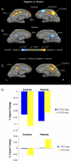
Fig. 1.
Fig. 1.
Negative Vs Neutral Contrast, medial cortical surface. Mean activation maps showing significant clusters of activation (P < .05) for the healthy control (n = 18) (A) and schizophrenia (n = 14) (B) group and the map of the between-group differences (C) for the negative vs. neutral contrast. For the within-group maps (A and B), warm colors indicate clusters showing greater responses to the negative relative to the neutral sentence pairs, while cold colors indicate clusters with greater activation to the neutral relative to the negative sentence pairs. For the map of the between-group differences (C), warm colors indicate clusters showing greater activation for the negative > neutral contrast in the control group compared with the patient group. (In C, there were no clusters showing greater activation in the patients compared with the controls for the negative > neutral contrast.) In D, percent signal change relative to baseline for the foci exhibiting significant between-group differences in C are displayed; the baseline is mean signal intensity. PCG = posterior cingulate gyrus, MFG = medial frontal gyrus, ACG = anterior cingulate gyrus, SFG = superior frontal gyrus, PrC = precuneus.
Fig. 2.
Fig. 2.
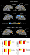
Positive Vs Neutral Contrast, medial cortical surface. Mean activation maps showing significant clusters of activation (P < .05) for the healthy control (n = 18) (A) and schizophrenia (n = 14) (B) group and the map of the between-group differences (C) for the positive vs neutral contrast. For the within-group maps (A and B), warm colors indicate clusters showing greater responses to the positive relative to the neutral sentence pairs, while cold colors indicate clusters with greater activation to the neutral relative to the positive sentence pairs. For the map of the between-group differences (C), warm colors indicate clusters showing greater activation for the positive > neutral contrast in the control group compared with the patient group. (In C, there were no clusters showing greater activation in the patients compared with the controls for the positive > neutral contrast.) In D, percent signal change relative to baseline for the foci exhibiting significant between-group differences in C are displayed; the baseline is mean signal intensity. PCG = posterior cingulate gyrus, MFG = medial frontal gyrus, ACG = anterior cingulate gyrus, SFG = superior frontal gyrus, PrC = precuneus, ACOC = anterior cingulate and orbital cortices.
Fig. 3.
Fig. 3.
Negative Vs Positive Contrast, Medial Cortical Surface. Mean activation maps, displayed on the medial cortical surface, showing significant clusters of activation (P < .05) for the healthy control (n = 18) (A) and schizophrenia (n = 14) (B) group and the map of the between-group differences (C) for the negative vs positive contrast. For the within-group maps (A and B), warm colors indicate clusters showing greater responses to the negative relative to the positive sentence pairs, while cold colors indicate clusters with greater activation to the positive relative to the negative sentence pairs. For the map of the between-group differences (C), cold colors indicate clusters showing greater activation for the negative > positive contrast in the patient group compared with the healthy control group. (In C, there were no clusters showing greater activation for the controls relative to the patients for the negative > positive contrast.) In D, percent signal change relative to baseline for the foci exhibiting significant between-group differences in C are displayed; the baseline is mean signal intensity. PCG = posterior cingulate gyrus, PrC = precuneus, MFG = medial frontal gyrus, ACG = anterior cingulate gyrus, ACOC = anterior cingulate and orbital cortices.
Fig. 4.
Fig. 4.
Correlation Between Delusion Severity and Cingulate Gyrus Response to the Neutral Stimuli in the Schizophrenia Patients. Graphs of the correlations between right (A) and left (B) cingulate gyrus responses (% signal change) to the neutral sentence pairs and delusion severity, measured using the SAPS global delusion item, in the 14 schizophrenia patients.

References

    1. Aleman A, Kahn RS. Strange feelings: do amygdala abnormalities dysregulate the emotional brain in schizophrenia? Prog Neurobiol. 2005;77:283–298. - PubMed
    1. Pinkham AE, Penn DL, Perkins DO, Lieberman J. Implications for the neural basis of social cognition for the study of schizophrenia. Am J Psychiatry. 2003;160:815–824. - PubMed
    1. Green MF, Leitman DI. Social cognition in schizophrenia. Schizophr Bull. 2008;34:670–672. - PMC - PubMed
    1. Poole JH, Tobias FC, Vinogradov S. The functional relevance of affect recognition errors in schizophrenia. J Int Neuropsychol Soc. 2000;6:649–658. - PubMed
    1. Phillips ML, Drevets WC, Rauch SL, Lane R. Neurobiology of emotion perception II: implications for major psychiatric disorders. Biol Psychiatry. 2003;54:515–528. - PubMed

Publication types