. 2009 Nov;81(5):978-88.
doi: 10.1095/biolreprod.109.078097. Epub 2009 Jul 15.

Neutralization of vascular endothelial growth factor antiangiogenic isoforms is more effective than treatment with proangiogenic isoforms in stimulating vascular development and follicle progression in the perinatal rat ovary

Affiliations

Neutralization of vascular endothelial growth factor antiangiogenic isoforms is more effective than treatment with proangiogenic isoforms in stimulating vascular development and follicle progression in the perinatal rat ovary

Robin A Artac et al. Biol Reprod. 2009 Nov.

Abstract

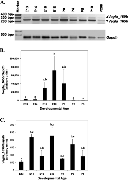
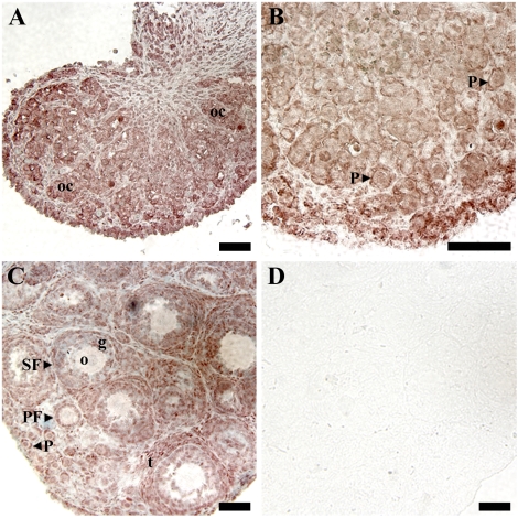
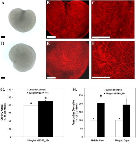
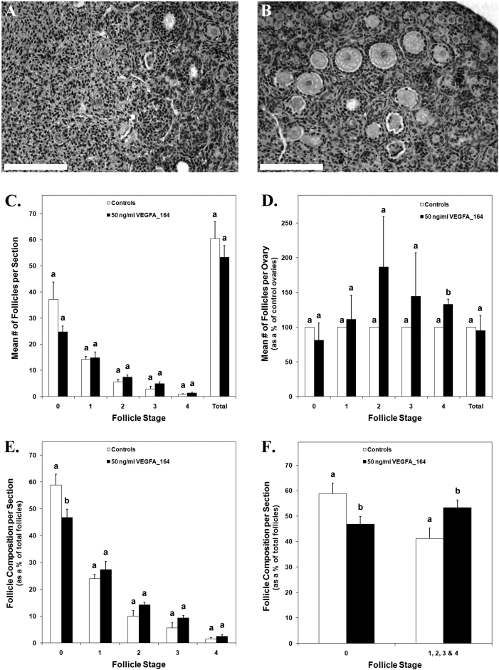
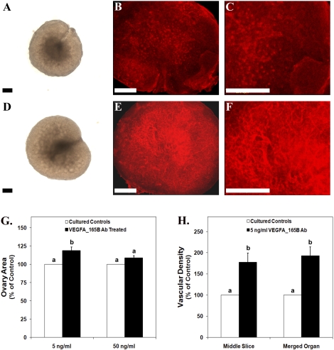
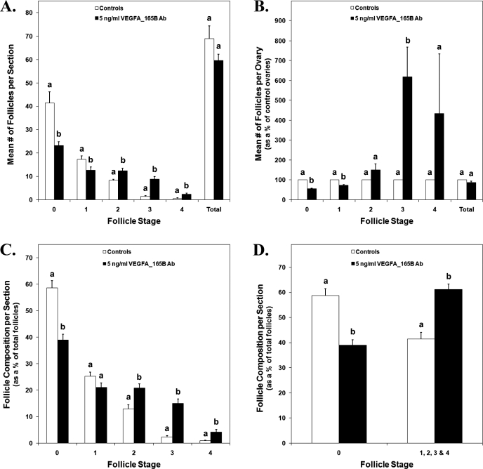
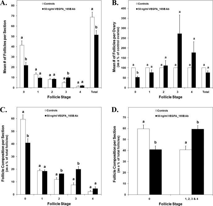
Inhibition of vascular endothelial growth factor A (VEGFA) signal transduction arrests vascular and follicle development. Because antiangiogenic VEGFA isoforms are proposed to block proangiogenic VEGFA isoforms from binding to their receptors, we hypothesized that proangiogenic isoforms promote and antiangiogenic isoforms inhibit these processes. The antiangiogenic isoforms Vegfa_165b and Vegfa_189b were amplified and sequenced from rat ovaries. The Vegfa_165b sequence was 90% homologous to human VEGFA_165B. Quantitative RT-PCR determined that Vegfa_165b mRNA was more abundant around Embryonic Day 18, but Vegfa_189b lacked a distinct pattern of abundance. Antiangiogenic VEGFA isoforms were localized to pregranulosa and granulosa cells of all follicle stages and to theca cells of advanced-stage follicles. To determine the effects of VEGFA isoforms in developing ovaries, Postnatal Day 3/4 rat ovaries were cultured with VEGFA_164 or an antibody to antiangiogenic isoforms (anti-VEGFAxxxB). Treatment with 50 ng/ml of VEGFA_164 resulted in a 93% increase in vascular density (P < 0.01), and treated ovaries were composed of fewer primordial follicles (stage 0) and more developing follicles (stages 1-4) than controls (P < 0.04). Ovaries treated with 5 ng/ml of VEGFAxxxB antibody had a 93% increase in vascular density (P < 0.02), with fewer primordial and early primary follicles (stage 1) and more primary, transitional, and secondary follicles (stages 2, 3, and 4, respectively) compared with controls (P < 0.005). We conclude that neutralization of antiangiogenic VEGFA isoforms may be a more effective mechanism of enhancing vascular and follicular development in perinatal rat ovaries than treatment with the proangiogenic isoform VEGFA_164.


Figures

FIG. 1.
FIG. 1.
A) Conventional RT-PCR for Vegfa antiangiogenic isoforms from E13 to P10 developing ovaries and P200 adult ovaries. Gapdh served as a control for RNA isolation and amplification. Negative control samples (without template) did not produce a band (data not shown). B and C) Quantitative RT-PCR to detect Vegfa_165b and Vegfa_189b from E13 to P5 of ovarian development. Gapdh was used as an endogenous control to account for differences in starting material. These data are the result of at least three different pools of tissue from each age group. The mean ± SEM normalized values are presented, and different letters represent a statistically significant difference in means (P < 0.05).
FIG. 2.
FIG. 2.
Immunohistochemistry for VEGFA antiangiogenic (xxxB) isoforms in P0 (A), P3 (B), and P10 (C and D) ovaries. A and C were lightly counterstained with hematoxylin. D) Postnatal Day 10 ovarian sections with no primary antibody served as negative controls. These data are the result of at least three different ovaries from each developmental time point. Bar = 50 μm. oc, oocyte cyst; P, primordial follicle; PF, primary follicle; SF, secondary follicle; o, oocyte; g, granulosa cells; t, theca cells.
FIG. 3.
FIG. 3.
Postnatal Day 3/4 ovarian organ cultures treated with vehicle control (AC) or 50 ng/ml of recombinant VEGFA_164 (DF). A and D) Bright-field images. B, C, E, and F) Confocal images of whole-mount immunohistochemistry staining for PECAM1 (red indicates endothelial cell marker) to localize vasculature. G) Effect of VEGFA_164 treatment on ovarian area expressed as a percentage of control organs. H) Effect of VEGFA_164 on vascular density (middle organ slice and total merged organ) expressed as a percentage of control organs. Bar = 150 μm. Data are the result of 12 (G) and 23 (H) ovary pairs. G and H) The mean ± SEM areas are presented, and different letters represent a statistically significant difference between treated and control groups (P < 0.01).
FIG. 4.
FIG. 4.
Postnatal Day 3/4 cultured control ovaries (A) or ovaries treated with 50 ng/ml of recombinant VEGFA_164 (B) stained with hematoxylin-eosin. C) The mean number of follicles per section from control and VEGFA_164 ovary pairs. D) The mean number of follicles for VEGFA_164-treated ovaries expressed as a percentage of their paired controls. E and F) The mean number of follicles per section for control and treated ovaries expressed as a percentage of the total number of follicles per section. Bar = 150 μm. CF) Data are the result of three ovary pairs and three histological sections evaluated at 200× magnification per ovary. The mean ± SEM numbers of follicles are presented, and different letters within each follicle stage represent a statistically significant difference (P < 0.04).
FIG. 5.
FIG. 5.
Postnatal Day 3/4 ovarian organ cultures treated with vehicle control (AC) or 5 ng/ml of VEGFAxxxB antibody (DF). A and D) Bright-field images. B, C, E, and F) Confocal images of whole-mount immunohistochemistry staining for PECAM1 (red indicates endothelial cell marker) to localize vasculature. G) Effect of VEGFAxxxB antibody treatment on ovarian area expressed as a percentage of control organs. H) Effect of 5 ng/ml of VEGFAxxxB antibody on vascular density (middle organ slice and total merged organ) expressed as a percentage of control organs. Bar = 150 μm. Data are the result of 12 (G [5 ng/ml]), 15 (G [50 ng/ml]), and 11 (H) ovary pairs. G and H) The mean ± SEM areas are presented, and different letters represent a statistically significant difference between treated and control groups (P < 0.02). Ab, antibody.
FIG. 6.
FIG. 6.
A) The mean number of follicles per section from control and 5 ng/ml of VEGFAxxxB antibody-treated P3/4 cultured ovary pairs. B) The mean number of follicles for VEGFAxxxB antibody-treated ovaries expressed as a percentage of their paired controls. C and D) The mean number of follicles per section for control and treated ovaries expressed as a percentage of the total number of follicles per section. AD) Data are the result of three ovary pairs and three histological sections evaluated at 200× magnification per ovary. The mean ± SEM numbers of follicles are presented, and different letters within each follicle stage represent a statistically significant difference (P < 0.05). Ab, antibody.
FIG. 7.
FIG. 7.
A) The mean number of follicles per section from control and 50 ng/ml VEGFAxxxB antibody-treated P3/4 cultured ovary pairs. B) The mean number of follicles for VEGFAxxxB antibody-treated ovaries expressed as a percentage of their paired controls. C and D) The mean number of follicles per section for control and treated ovaries expressed as a percentage of the total number of follicles per section. AD) Data are the result of three ovary pairs and three histological sections evaluated at 200× magnification per ovary. The mean ± SEM numbers of follicles are presented, and different letters within each follicle stage represent a statistically significant difference (P < 0.03). Ab, antibody.

References

    1. Hirshfield AN.Heterogeneity of cell populations that contribute to the formation of primordial follicles in rats. Biol Reprod 1992; 47: 466–472.. - PubMed
    1. Rajah R, Glaser EM, Hirshfield AN.The changing architecture of the neonatal rat ovary during histogenesis. Dev Dyn 1992; 194: 177–192.. - PubMed
    1. Pepling ME, Spradling AC.Mouse ovarian germ cell cysts undergo programmed breakdown to form primordial follicles. Dev Biol 2001; 234: 339–351.. - PubMed
    1. Nilsson E, Skinner MK.Cellular interactions that control primordial follicle development and folliculogenesis. J Soc Gynecol Investig 2001; 8: S17–S20.. - PubMed
    1. Parrott JA, Skinner MK.Kit-ligand/stem cell factor induces primordial follicle development and initiates folliculogenesis. Endocrinology 1999; 140: 4262–4271.. - PubMed

Publication types

MeSH terms