Review
. 2009 Aug 7;9(15):2132-9.
doi: 10.1039/b903043c. Epub 2009 Jun 4.

Biological implications of polydimethylsiloxane-based microfluidic cell culture

Affiliations
Review

Biological implications of polydimethylsiloxane-based microfluidic cell culture

Keil J Regehr et al. Lab Chip. .

Abstract

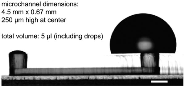
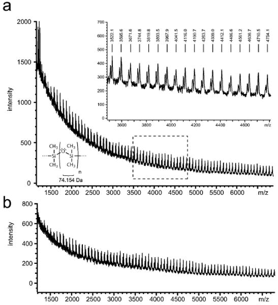
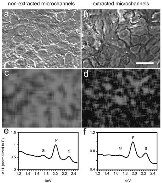
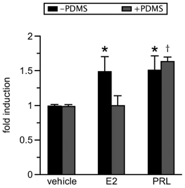
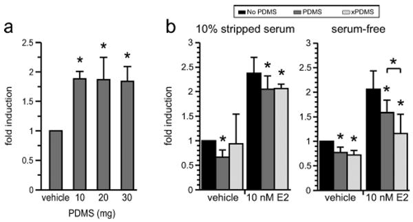
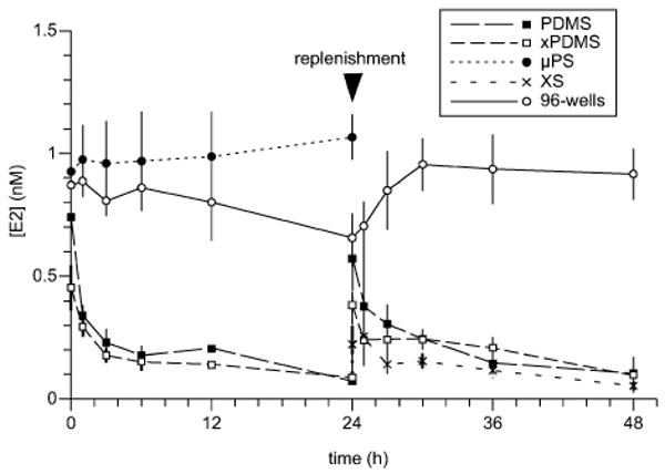
Polydimethylsiloxane (PDMS) has become a staple of the microfluidics community by virtue of its simple fabrication process and material attributes, such as gas permeability, optical transparency, and flexibility. As microfluidic systems are put toward biological problems and increasingly utilized as cell culture platforms, the material properties of PDMS must be considered in a biological context. Two properties of PDMS were addressed in this study: the leaching of uncured oligomers from the polymer network into microchannel media, and the absorption of small, hydrophobic molecules (i.e. estrogen) from serum-containing media into the polymer bulk. Uncured PDMS oligomers were detectable via MALDI-MS in microchannel media both before and after Soxhlet extraction of PDMS devices in ethanol. Additionally, PDMS oligomers were identified in the plasma membranes of NMuMG cells cultured in PDMS microchannels for 24 hours. Cells cultured in extracted microchannels also contained a detectable amount of uncured PDMS. It was shown that MCF-7 cells seeded directly on PDMS inserts were responsive to hydrophilic prolactin but not hydrophobic estrogen, reflecting its specificity for absorbing small, hydrophobic molecules; and the presence of PDMS floating in wells significantly reduced cellular response to estrogen in a serum-dependent manner. Quantification of estrogen via ELISA revealed that microchannel estrogen partitioned rapidly into the surrounding PDMS to a ratio of approximately 9:1. Pretreatments such as blocking with serum or pre-absorbing estrogen for 24 hours did not affect estrogen loss from PDMS-based microchannels. These findings highlight the importance of careful consideration of culture system properties when determining an appropriate environment for biological experiments.


Figures

Fig. 1
Fig. 1
A PDMS-based microfluidic channel of the type used in this study.
Fig. 2
Fig. 2
PDMS oligomers are detectable in microchannel media. (a) Uncured PDMS oligomers were detectable via MALDI-MS in DIUF water aspirated from microchannels after a 24-hour incubation. (b) Uncrosslinked oligomers were still detected in water aspirated from Soxhlet-extracted microchannels.
Fig. 3
Fig. 3
PDMS oligomers are detectable in cell membranes. SEM of NMuMG luminal cells cultured in non-extracted PDMS (a) or Soxhlet-extracted PDMS (b) microchannels, with corresponding EDX maps of silicon content over the same regions of interest (c, d). Silicon was profusely distributed in the plasma membranes of cells cultured in non-extracted PDMS microchannels for 24 h. The silicon content was substantially reduced but detectable in the membranes of cells cultured in extracted microchannels. Spectra showing silicon, phosphorus, and sulfur peaks for both cases represent full-image regions of interest (e, f). Scale bar = 20 μm.
Fig. 4
Fig. 4
PDMS selectively inhibits estrogen signaling. MCF-7 cells were transiently transfected with a 4xAP-1-luciferase construct and seeded in 12-well plates with or without PDMS inserts. Cells in direct contact with PDMS (seeded on PDMS inserts) (+PDMS) displayed significant AP-1 activity in response to prolactin (PRL, 4 nM) but not estrogen (E2, 1 nM). Cells cultured in the absence of PDMS (−PDMS) displayed significant AP-1 activity in response to both estrogen and PRL (n = 3 experiments). *p < 0.05; p < 0.001.
Fig. 5
Fig. 5
PDMS absorbs and releases estrogen. PDMS exhibits capacitance for small, hydrophobic molecules. (a) Pieces of PDMS of mass 10, 20, or 30 mg were incubated individually for 24 hours in 100 μl of serum-free media containing 10−7 M E2. When thoroughly rinsed and transferred to wells containing cells in stripped serum, estrogen was released from the PDMS in a manner independent of the mass, indicating that the PDMS was not saturated with estrogen (p < 0.005 each, n = 3 experiments). (b) MCF-7 MVLN cells stably expressing luciferase were cultured in 10% stripped serum or serum-free media in the presence of no PDMS (No PDMS), non-extracted PDMS (PDMS), or extracted PDMS (xPDMS) pieces. Left, Cells cultured in stripped serum exhibited no difference in luciferase induction between PDMS treatments, though both conditions caused a statistically significant drop in induction from no-PDMS controls. Right, Cells cultured in the absence of serum showed a clearer distinction between non-extracted and extracted PDMS in response to estrogen. Both PDMS conditions effected a statistically significant inhibition of estrogen signaling, and at 10−8 M estrogen were significantly different from each other (p < 0.05, n = 3 experiments).
Fig. 6
Fig. 6
Time-dependent estrogen loss from microchannel media. Estrogen was lost to the surrounding PDMS in all PDMS-based microchannels, irrespective of pretreatment. Microchannels made from either non-extracted PDMS (PDMS) or Soxhlet-extracted PDMS (xPDMS) lost estrogen readily in the first 24 hours after filling with 1 nM estrogen-supplemented, phenol red-free DMEM. Other extracted channels were first incubated for 24 hours with estrogen-supplemented DMEM (xPDMS) or 10% stripped serum (XS) and sampled over the following 24 hours. Neither pretreatment was able to prevent estrogen absorption into the bulk during the second 24 hours. Polystyrene wells (96-wells) and tissue-culture treated polystyrene microchannels (μPS) did not lose estrogen progressively over time}.

References

    1. Kane R, Takayama S, Ostuni E, Ingber D, Whitesides G. Biomaterials. 1999;20:2363–2376. - PubMed
    1. Jo B, Van Lerberghe L, Motsegood K, Beebe D. J Microelectomech Syst. 2000;9:76–81.
    1. Leclerc E, Sakai Y, Fujii T. Biomedical Microdevices. 2003;5:109–114.
    1. Nikkhah M, Strobl J, Agah M. Proc IEEE EMBS. 2007;29:6077–6080.
    1. Gross P, Kartalov E, Scherer A, Weiner L. J Neurol Sci. 2007;252:135–143. - PubMed

Publication types

MeSH terms