. 2009 Sep 11;392(1):208-17.
doi: 10.1016/j.jmb.2009.07.018. Epub 2009 Jul 14.

Prokaryotic ubiquitin-like protein pup is intrinsically disordered

Affiliations

Prokaryotic ubiquitin-like protein pup is intrinsically disordered

Xiang Chen et al. J Mol Biol. .

Abstract

The prokaryotic ubiquitin-like protein Pup targets substrates for degradation by the Mycobacterium tuberculosis proteasome through its interaction with Mpa, an ATPase that is thought to abut the 20S catalytic subunit. Ubiquitin, which is assembled into a polymer to similarly signal for proteasomal degradation in eukaryotes, adopts a stable and compact structural fold that is adapted into other proteins for diverse biological functions. We used NMR spectroscopy to demonstrate that, unlike ubiquitin, the 64-amino-acid protein Pup is intrinsically disordered with small helical propensity in the C-terminal region. We found that the Pup:Mpa interaction involves an extensive contact surface that spans S21-K61 and that the binding is in the "slow exchange" regime on the NMR time scale, thus demonstrating higher affinity than most ubiquitin:ubiquitin receptor pairs. Interestingly, during the titration experiment, intermediate Pup species were observable, suggesting the formation of one or more transient state(s) upon binding. Moreover, Mpa selected one configuration for a region undergoing chemical exchange in the free protein. These findings provide mechanistic insights into Pup's functional role as a degradation signal.


Figures
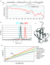
Figure 1
Figure 1. Prokaryotic ubiquitin-like protein Pup is rich in low complexity sequence and has a larger than expected hydrodynamic radius under native conditions
(a) The amino acid sequence of Pup is displayed with its aspartic and glutamic acids in grey and its diglycine motif and C-terminal glutamine in red and blue, respectively. Its predicted secondary structure (with the program Jpred33) and sequence complexity (with the program seg34) is displayed below the sequence. Helical regions and sequences of low complexity are displayed with ‘H’ and an ‘X,’ respectively. The results of the disorder prediction programs PONDR VSL2 and IUPred are plotted as labeled; scores over 0.5 indicate predicted disorder. (b) Size exclusion chromatrography by FPLC reveals that 6.9 kDa Pup has a larger hydrodynamic radius than 8.6 kDa ubiquitin. At the bottom of the chromatogram, we indicate the reported elution volume for ferritin, albumin, ovalbumin and myoglobin with arrows labeled a, b, c, and d, respectively. (c) A ribbon diagram is displayed of ubiquitin to highlight its structural complexity. Ubiquitin targets proteins for degradation in eukaryotes, but unlike Pup forms a well-folded, compact structure. (d) Pup’s apparent Rs based on its elution time in (b) matches that of a natively unfolded protein. Stokes radius, Rs; MW, molecular weight.
Figure 2
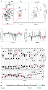
Figure 2. NMR data indicate that Pup contains a stable helix spanning A51–F54
(a) 1H, 15N HSQC experiments reveal that Pup (shown on the left) lacks the amide chemical shift dispersion characteristic of a folded protein (as displayed for ubiquitin on the right). R29, V55–V59 exhibited two sets of amide crosspeaks, as labelled. (b) Chemical shift index (CSI) profile for Pup Cα (left) and Hα (right) atoms are displayed. Residues A51–F54 are highlighted in red, as they demonstrate the shifting characteristic of helical structures and were also demonstrated to be helical in an 15N dispersed NOESY spectrum (Figure c). CSI values were determined by subtracting random coil chemical shift value from the assigned one. (c) Regions with long-range NOE interactions are displayed for an 15N dispersed NOESY spectrum acquired on 15N labeled Pup in 20 mM HEPES (pH 6.5) and 50 mM NaCl; no glycerol was used for this experiment. Intra-residue, sequential, i → i+2, and i → i+3 interactions are displayed in black, red, purple, and blue, respectively. (d) Summary of NOESY and CSI data demonstrating that residues A51–F54 (highlighted in blue) exhibit a propensity towards helicity while the rest of the sequence is largely devoid of canonical secondary structure elements. Residues undergoing slow conformational exchange are indicated in grey.
Figure 3
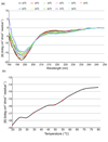
Figure 3. Circular dichroism (CD) data demonstrate that Pup does not unfold cooperatively
(a) CD spectra in the far-UV region are monitored at several temperatures spanning from 20 to 80 °C on Pup by using a spectropolarimeter equipped with a water circulation temperature control. (b) Thermal unfolding transition curves are provided by plotting the change in ellipticity at 197.7 nm from (a) across temperature. No melting transition is observed for Pup.
Figure 4
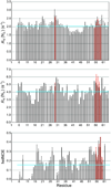
Figure 4. NMR relaxation experiments demonstrate that Pup is a dynamic protein with regions of conformational exchange
(a) Amide longitudinal (RN(NZ)) and (b) transverse (RN(NX)) relaxation rates and (c) 15N heteronuclear NOE enhancements (hetNOE) are displayed for Pup. Both sets of resonances are displayed for R29 and V55–V59 with the arbitrarily assigned “second” resonance displayed in red. The average value is displayed with a blue line to highlight sequence variations.
Figure 5
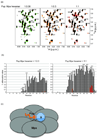
Figure 5. The C-terminal region of Pup binds to the Mycobacterium proteasomal ATPase Mpa
(a) 1H, 15N HSQC titration experiment in which unlabeled Mpa is added to 15N labeled Pup. Free Pup (black) is displayed superimposed onto Pup:Mpa hexamer at molar ratios of 1:0.08, 1:0.3, and 1:1 as indicated. Resonances from the Mpa-bound state that persist throughout the titration are highlighted with an asterisk, whereas those that appear and then disappear are marked with an arrow. The binding is in slow exchange and therefore the Mpa-bound state is not assigned. (b) Quantitative analysis of the Pup residues involved in Mpa hexamer binding plotting 1attenuation=1(I1:0.3Ifree) (left panel) or 1(I1:1Ifree) (right panel) for each residue in the Pup sequence. In this equation, I1:0.3 and I1:1 is the intensity of the unbound Pup resonance with 0.3 and equimolar ratio of Mpa hexamer; Ifree is the intensity of the Pup resonance with no Mpa hexamer present. (c) A model depicting a pupylated substrate binding to Mpa, which forms a hexamer. Pup, its substrate, and Mpa are displayed in orange, blue and grey, respectively.

References

    1. Zwickl P, Baumeister W, Steven A. Dis-assembly lines: the proteasome and related ATPase-assisted proteases. Curr Opin Struct Biol. 2000;10:242–250. - PubMed
    1. Nagy I, Tamura T, Vanderleyden J, Baumeister W, De Mot R. The 20S proteasome of Streptomyces coelicolor. J Bacteriol. 1998;180:5448–5453. - PMC - PubMed
    1. Pouch MN, Cournoyer B, Baumeister W. Characterization of the 20S proteasome from the actinomycete Frankia. Mol Microbiol. 2000;35:368–377. - PubMed
    1. Darwin KH, Ehrt S, Gutierrez-Ramos JC, Weich N, Nathan CF. The proteasome of Mycobacterium tuberculosis is required for resistance to nitric oxide. Science. 2003;302:1963–1966. - PubMed
    1. Knipfer N, Shrader TE. Inactivation of the 20S proteasome in Mycobacterium smegmatis. Mol Microbiol. 1997;25:375–383. - PubMed

Publication types

MeSH terms

Substances

LinkOut - more resources