Review
. 2009 Aug;8(8):731-40.
doi: 10.1016/S1474-4422(09)70134-6.

Varicella zoster virus vasculopathies: diverse clinical manifestations, laboratory features, pathogenesis, and treatment

Affiliations
Review

Varicella zoster virus vasculopathies: diverse clinical manifestations, laboratory features, pathogenesis, and treatment

Don Gilden et al. Lancet Neurol. 2009 Aug.

Abstract

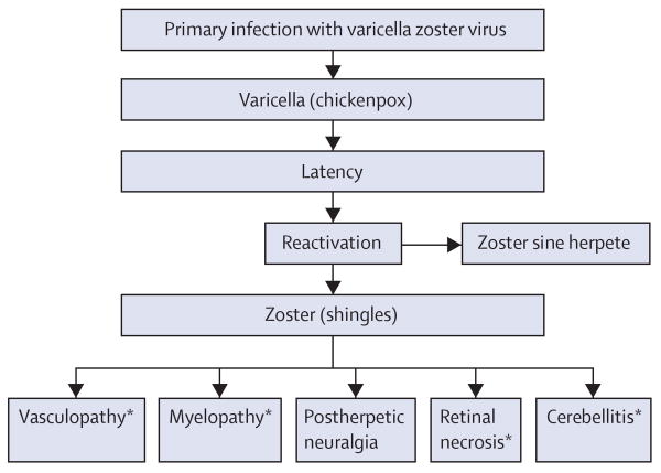
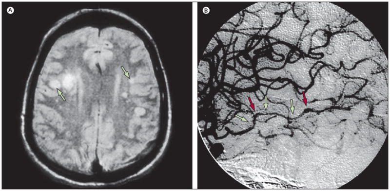
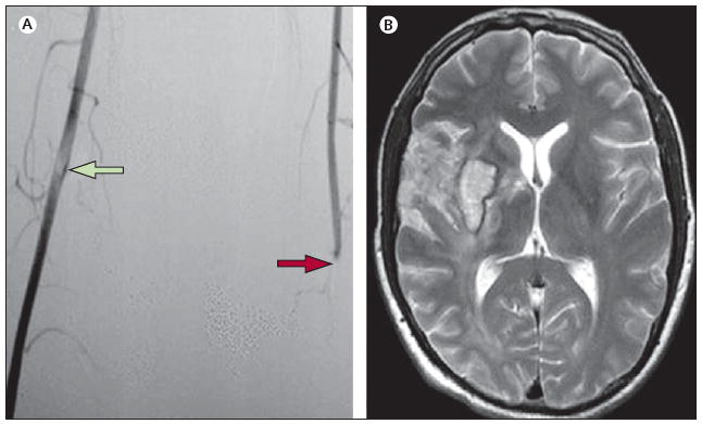
Vasculopathies caused by varicella zoster virus (VZV) are indicative of a productive virus infection in cerebral arteries after either reactivation of VZV (shingles) or primary infection (chickenpox). VZV vasculopathy can cause ischaemic infarction of the brain and spinal cord, as well as aneurysm, subarachnoid and cerebral haemorrhage, carotid dissection, and, rarely, peripheral arterial disease. VZV vasculopathy in immunocompetent or immunocompromised individuals can be unifocal or multifocal with deep-seated and superficial infarctions. Lesions at the grey-white matter junction on brain imaging are a clue to diagnosis. Involvement of both large and small arteries is more common than that of either alone. Most patients have a mononuclear cerebrospinal fluid pleocytosis, often with red blood cells. Cerebrospinal fluid pleocytosis and rash are absent in about a third of cases. Anti-VZV IgG antibody in the cerebrospinal fluid is found more frequently than VZV DNA. In recent years, the number of recognised VZV vasculopathies has grown, and accurate diagnosis is important for the effective treatment of these disorders.


Conflict of interest statement

Conflicts of interest: We have no conflicts of interest.

Figures

Figure 1
Figure 1. Neurological disease caused by reactivation of varicella zoster virus
*Can occur after varicella and can also occur without a rash.
Figure 2
Figure 2. MRI scan and cerebral angiogram of patients with VZV vasculopathies
(A) MRI scan of a patient with VZV multifocal vasculopathy. Proton-density brain MRI scan shows multiple areas of infarction in both hemispheres, particularly involving the white matter. Arrows point to lesions at the grey–white matter junctions. Reproduced from Gilden and co-workers, with permission from Taylor & Francis. (B) Cerebral angiogram from a patient with VZV vasculopathy. Focal areas of stenosis (green arrows) and poststenotic dilatation (red arrows) involving the right posterior cerebral artery can be seen (left side of picture, right side of patient). Reproduced from Russman and co-workers, with permission from the American Medical Association. VZV=varicella zoster virus.
Figure 3
Figure 3. Aneurysm and subarachnoid haemorrhage in patients with VZV vasculopathy
(A) Aneurysm in a patient with VZV vasculopathy. Angiogram 4 weeks after zoster reveals an aneurysm in the intrapetrosal portion of the left internal carotid artery (arrow). Reproduced from Gürsoy and co-workers, with permission from Springer-Verlag. (B) Subarachnoid haemorrhage in a patient with zoster and VZV vasculopathy. Axial CT shows subarachnoid haemorrhage and a left para-midline haematoma (left side of picture, right side of patient). Reproduced from Jain and co-workers, with permission from the American Society of Neuroradiology. VZV=varicella zoster virus.
Figure 4
Figure 4. VZV vasculopathy affecting cerebral and peripheral arteries
(A) Lower-limb angiography shows occlusion of the left femoral artery (red arrow) and the presence of luminal non-occlusive thrombi in the right femoral artery (green arrow). (B) Axial T2-weighted brain MRI of the same patient revealed an ischaemic lesion involving the right striatocapsular region, adjacent white matter, and frontotemporal cortex laterally and superiorly. Small haemorrhagic foci were seen inside the ischaemic lesion; magnetic resonance angiogram revealed proximal occlusion of the right middle cerebral artery (not shown). No other pathological findings were apparent (left side of picture, right side of patient). Reproduced from Massano and co-workers, with permission from John Wiley. VZV=varicella zoster virus.
Figure 5
Figure 5. Pathological and virological findings in arteries of patients who died from VZV vasculopathy
(A) VZV DNA in the posterior cerebral artery (lane 3) and basilar artery (lane 5). (B) VZV antigen (red) in the media of a cerebral artery. Panels A and B are reproduced from Gilden and co-workers, with permission from the American Academy of Neurology. (C) Cerebral artery with multinucleated giant cells (arrow). (D) Multiple herpes virions within a cerebral artery. CMV=cytomegalovirus. HSV=herpes zoster virus 1. VZV=varicella zoster virus.

References

    1. Askalan R, Laughlin S, Mayank S, et al. Chicken pox and stroke in childhood. A study of frequency and causation. Stroke. 2001;32:1257–62. - PubMed
    1. Braun KPJ, Bulder MMM, Chabrier S, et al. The course and outcome of unilateral intracranial arteriopathy in 79 children with ischaemic stroke. Brain. 2009;132:544–57. - PMC - PubMed
    1. Cravioto H, Feigin I. Noninfectious granulomatous angiitis with a predilection for the nervous system. Neurology. 1959;9:599–608. - PubMed
    1. Rosenblum WI, Hadfield MG. Granulomatous angiitis of the nervous system in cases of herpes zoster and lymphosarcoma. Neurology. 1972;22:348–54. - PubMed
    1. Gilbert GJ. Herpes zoster ophthalmicus and delayed contralateral hemiparesis. Relationships of the syndrome in central nervous system granulomatous angiitis. JAMA. 1974;229:302–04. - PubMed