Comparative Study
. 2009 Dec;50(12):5778-84.
doi: 10.1167/iovs.09-3790. Epub 2009 Jul 15.

Automated segmentation of the cup and rim from spectral domain OCT of the optic nerve head

Affiliations
Comparative Study

Automated segmentation of the cup and rim from spectral domain OCT of the optic nerve head

Michael D Abràmoff et al. Invest Ophthalmol Vis Sci. 2009 Dec.

Abstract

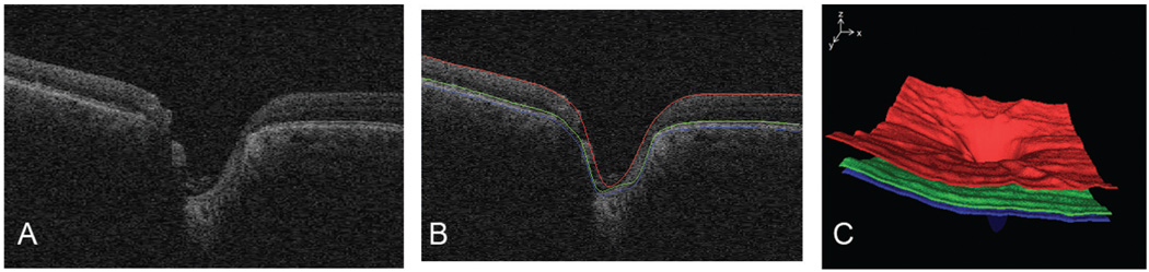
Purpose: To evaluate the performance of an automated algorithm for determination of the cup and rim from close-to-isotropic spectral domain (SD) OCT images of the optic nerve head (ONH) and compare to the cup and rim as determined by glaucoma experts from stereo color photographs of the same eye.

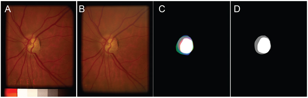
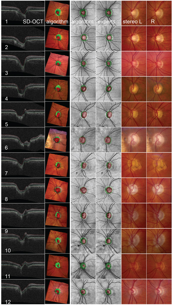
Methods: Thirty-four consecutive patients with glaucoma were included in the study, and the ONH in the left eye was imaged with SD-OCT and stereo color photography on the same day. The cup and rim were segmented in all ONH OCT volumes by a novel voxel column classification algorithm, and linear cup-to-disc (c/d) ratio was determined. Three fellowship-trained glaucoma specialists performed planimetry on the stereo color photographs, and c/d was also determined. The primary outcome measure was the correlation between algorithm-determined c/d and planimetry-derived c/d.

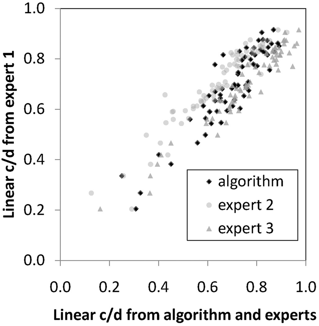
Results: The correlation of algorithm c/d to experts 1, 2, and 3 was 0.90, 0.87, and 0.93, respectively. The c/d correlation of expert 1 to 2, 1 to 3, and 2 to 3, were 0.89, 0.93, and 0.88, respectively.

Conclusions: In this preliminary study, we have developed a novel algorithm to determine the cup and rim in close-to-isotropic SD-OCT images of the ONH and have shown that its performance for determination of the cup and rim from SD-OCT images is similar to that of planimetry by glaucoma experts. Validation on a larger glaucoma sample as well as normal controls is warranted.


Figures

FIGURE 1
FIGURE 1
(A, B) Left and right image of a stereo pair of an optic disc, the original captured image was cropped and separated. The left and right stereo images can be fused by holding the page at about 30 cm (12 in.) from the eye and gazing into infinity. (C) Planimetry by three glaucoma specialists superimposed, to show how the reference standard in (D) is created. It shows the cup and rim of each expert in bright red, green, and blue (rim), respectively, pale red, green, and blue (cup). Thus, where all three experts indicated cup, white is shown, and where all three experts indicated rim, gray is shown. (D) Reference standard developed from the planimetries shown in (C).
FIGURE 2
FIGURE 2
Layer segmentation before flattening. (A) Single slice of SD-OCT scan of optic nerve head of the same patient and the same eye as in Figure 1. (B) Three surfaces segmented by using fast layer segmentation. (C) Surface rendering of these three surfaces.
FIGURE 3
FIGURE 3
Volume-flattening step. Left: after three surfaces have been detected, a 3-D thin plate spline is fitted to the surface, except through the area of the ONH, where this spline is extrapolated. Right: the entire volume is then reconstructed, also in 3-D, based on this spline fitted to surface 3.
FIGURE 4
FIGURE 4
Propagation of the reference standard from stereo color image–based planimetry to SD-OCT volume. (A) Left stereo color fundus image with superimposed reference standard (reddish cup, greenish rim). (B) Vessel segmentation in a surface projection of an SD-OCT image. (C) Co-localization of an SD-OCT projection image and left stereo color fundus image. (D) Propagation of cup (reddish) and rim (greenish) voxel columns from the reference standard in a stereo color fundus image into an SD-OCT image (single vertical slice shown) based on co-localization in (C).
FIGURE 5
FIGURE 5
The correlation of the algorithm-determined linear c/d with that of any glaucoma specialist was not inferior to the correlation of any glaucoma specialist to any other glaucoma specialist. Scatterplot of linear c/d of the algorithms presented in this paper and of glaucoma specialist 2 and glaucoma specialist 3, when the algorithm was trained on the reference standard for the training set only cup and disc segmentation only.
FIGURE 6
FIGURE 6
Visualization of the performance of automated segmentation of an optic nerve head (ONH) from SD-OCT for 12 of 34 randomly selected eyes, compared with the performance of three glaucoma specialists. Top to bottom rows 1–12: 12 patients, left eye. Left to right: SD-OCT: central slice 100/200 of the SD-OCT volume through the center of the disc, oriented from nasal (left) to temporal (right), with three detected surfaces indicated; algorithm: visualization of the detected cup (reddish) and rim (greenish) on a 3-D surface rendering of the ONH wrapped with color fundus photograph (the shape of the surface derived from the OCT and the color of the surface derived from the co-registered color image) to show the three-dimensional relationship of the cup and rim; algorithm: detected cup and rim on SD-OCT projection image; experts: propagation of the reference standard created by three glaucoma specialists from stereo color photos onto SD-OCT projection image; stereo L and R: left and right color stereo photos that experts used to obtain reference standard.

References

    1. Tielsch JM, Sommer A, Katz J, Royall RM, Quigley HA, Javitt J. Racial variations in the prevalence of primary open-angle glaucoma: The Baltimore Eye Survey. JAMA. 1991;266(3):369–374. - PubMed
    1. Heijl A, Leske MC, Bengtsson B, Hyman L, Bengtsson B, Hussein M. Reduction of intraocular pressure and glaucoma progression: results from the Early Manifest Glaucoma Trial. Arch Ophthalmol. 2002;120(10):1268–1279. - PubMed
    1. Greaney MJ, Hoffman DC, Garway-Heath DF, Nakla M, Coleman AL, Caprioli J. Comparison of optic nerve imaging methods to distinguish normal eyes from those with glaucoma. Invest Ophthalmol Vis Sci. 2002;43(1):140–145. - PubMed
    1. Tielsch JM, Katz J, Quigley HA, Miller NR, Sommer A. Intraobserver and interobserver agreement in measurement of optic disc characteristics. Ophthalmology. 1988;95(3):350–356. - PubMed
    1. Kwon Y, Adix M, Zimmerman MB, et al. Variance due to observer, repeat imaging, and fundus camera type on cup-to-disc ratio estimates by stereo planimetry. J Glaucoma. 2009;18(4):305–310. - PubMed

Publication types