. 2009 Nov;94(11):1493-501.
doi: 10.3324/haematol.2009.006072. Epub 2009 Jul 16.

The integrin alpha9beta1 on hematopoietic stem and progenitor cells: involvement in cell adhesion, proliferation and differentiation

Affiliations

The integrin alpha9beta1 on hematopoietic stem and progenitor cells: involvement in cell adhesion, proliferation and differentiation

Thomas D Schreiber et al. Haematologica. 2009 Nov.

Abstract

Background: Hematopoietic stem and progenitor cells can interact with their microenvironment via integrins which are adhesion receptors consisting of alpha and beta subunits. Current knowledge suggests that the integrin subunits alpha4 and alpha6 expressed on hematopoietic stem and progenitor cells have distinct roles in retaining stem cells in the bone marrow. The aim of our study was to gain insight into the expression and functions of the integrin subunits alpha7-alpha11 within the endosteal stem cell niche.

Design and methods: Human osteoblasts isolated from trabecular bone and hematopoietic stem and progenitor cells purified from umbilical cord blood or bone marrow aspirates were analyzed for the expression of integrin alpha7-alpha11 chains by reverse transcriptase polymerase chain reaction. The involvement of the integrin alpha9beta1 in hematopoietic stem and progenitor cell adhesion, proliferation and differentiation was analyzed in functional assays.

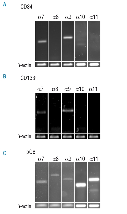
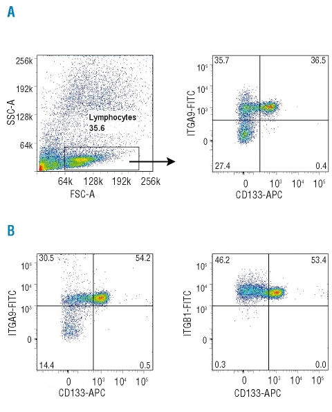
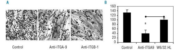
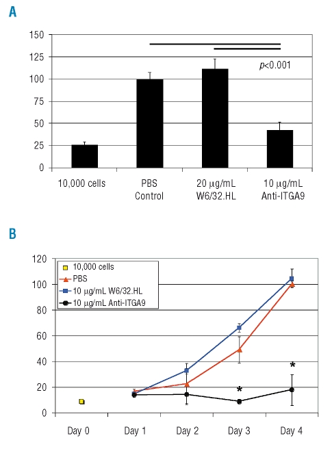
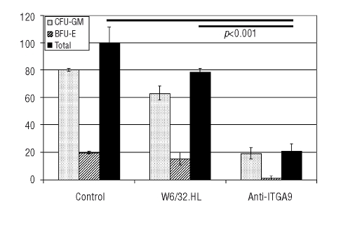
Results: Transcripts for all investigated integrin chains were found in primary osteoblasts. Highly purified hematopoietic stem and progenitor cells, however, expressed only transcripts encoding integrin subunits alpha7 and alpha9. Flow cytometric analysis verified extracellular expression of the integrin alpha9beta1 on hematopoietic stem and progenitor cells. Cell-cell adhesion assays with osteoblasts and dye-labeled CD34(+) hematopoietic stem and progenitor cells in the presence of function-blocking antibodies revealed a role of integrin alpha9 in hematopoietic stem and progenitor cell adhesion to osteoblasts. Furthermore, the addition of anti-integrin alpha9 antibodies significantly inhibited proliferation and in vitro differentiation of CD34(+) hematopoietic stem and progenitor cells.

Conclusions: The integrin alpha9beta1 has been identified as a new member of the integrin beta1-subfamily expressed on human hematopoietic stem and progenitor cells. The functional studies strongly suggest that integrin alpha9beta1 contributes to adhesion and differentiation of hematopoietic stem and progenitor cells in the endosteal stem cell niche.


Figures

Figure 1.
Figure 1.
RT-PCR analysis of integrin α7-α11 mRNA expression in human CD34+ cells, CD133+ cells and human primary osteoblasts. RNA of isolated cells was reverse-transcribed in cDNA and amplified with the indicated primers. β-actin was amplified as a positive control. Human CD34+ cells (A) and CD133+ cells (B), isolated from umbilical cord blood, expressed the integrin α subunits 7 and 9, whereas human primary osteoblasts (pOB) expressed all investigated integrin subunits α7 - α11 (C).
Figure 2.
Figure 2.
Expression of integrin α9β1 on human bone marrow and umbilical cord blood CD133+ cells. Bone marrow mononuclear cells or umbilical cord blood cells were depleted of cells expressing lineage markers. Lineage-negative cells were labeled with anti-integrin α9 antibodies and antibodies against the stem and progenitor cell marker CD133 and analyzed by flow cytometry. (A) The lymphocyte-containing fraction of lineage-negative cells isolated from human bone marrow was selected for further analysis (left); 36.5% of these selected cells expressed both CD133 and the integrin α9 chain, whereas only 0.4% expressed CD133 but not integrin α9, indicating that >98.5% of the CD133+ cells in the lymphocyte-containing fraction expressed the integrin α9 chain (right). (B) An analysis similar to that shown for bone marrow cells was also performed for human umbilical cord blood cells; 99% of the CD133+ cells of the lymphocyte-containing fraction expressed the integrin α9 (left), and analogously, 99.7% also expressed the integrin β1 chain (right).
Figure 3.
Figure 3.
Influence of integrin α9β1 on HSPC cell adhesion to primary osteoblasts. MACS-isolated CD34+ HSPC were allowed to adhere to a confluent layer of isolated primary osteoblasts. (A) Without addition of an antibody, the CD34+ cells (small dark balls) adhered strongly to the osteoblasts, which can be recognized as the cell layer underlying the CD34+ cells. Addition of the monoclonal antibodies against the integrin α9 chain or the integrin β1 chain drastically reduced the number of adherent CD34+ cells (crystal violet stain, Zeiss Axiovert, magnification x100). (B) Quantification with BCECF-labeled CD34+ cells revealed that adhesion of these cells was significantly (p<0.05) reduced to 40±15% of that seen in the experiment with the control antibody W6/32.HL, which binds to MHC class I molecules on the cell surface. No significant difference was observed between the control experiment without antibody treatment (control) and the treatment with the antibody W6/32.HL. The individual experiments were performed in triplicate, and means of four independent experiments are shown with standard error bars.
Figure 4.
Figure 4.
Cell attachment of human CD34+ HSPC to VCAM-1 and the extracellular matrix molecules tenascin-C and osteopontin. MACS-isolated CD34+ cells were allowed to attach to immobilized vascular cell adhesion molecule (VCAM-1), tenascin-C (TN-C) and osteopontin (OPN). Strong attachment was observed for VCAM-1 and tenascin-C, but not for osteopontin (dark field illumination, Zeiss Axiovert, magnification x60). Cell adhesion to VCAM-1 and tenascin-C was not inhibited by anti-α9-integrin antibodies (VCAM-1, α-Int-α9 and TN-C, α-Int-α9) or by the control antibody W6/32.HL (α-HL).
Figure 5.
Figure 5.
Inhibition of cell proliferation of human CD34+ cells by the anti-integrin-α9 antibody Y9A2. (A) Isolated human CD34+ HSPC (1×10⁴) were cultured for 4 days with the indicated antibodies in serum-free expansion medium including 20 ng/mL each of interleukin-6 and as well as 100 ng/mL each of stem cell factor and Flt3L. Cell proliferation was measured with the CyQuant Cell Proliferation Assay by fluorescence read-out. Addition of anti-integrin α9 antibodies (anti-ITGA9) reduced cell proliferation significantly compared to the addition of W6/32.HL antibodies or the condition without antibody treatment (PBS control). Means with standard error bars are shown. n independent experiments were performed in duplicate. n=4 for PBS and anti-ITGA9, n=3 for W6/32.HL. (B) Time course of HSPC proliferation in the presence of 10 μg/mL anti-integrin α9 antibody (Y9A2). Isolated human CD34+ HSPC (1×10⁴) were cultured for 4 days as described above. Cell proliferation was analyzed each day with the CyQuant Cell Proliferation Assay. Up to day 4, CD34+ cells incubated in the presence of W6/32.HL antibodies or without antibodies proliferated approximately five times better than cells cultured with anti-integrin α9 antibodies, which only doubled in number compared to the input. One representative experiment out of three independent experiments is shown. Means of duplicates with standard deviation are shown. *=p<0.05. RFU: relative fluorescence units.
Figure 6.
Figure 6.
Colony formation assay of human CD34+ cells in the presence of 5 μg anti-integrin α9 antibody (Y9A2). Isolated human CD34+ HSPC (1.6×10³) were incubated for 14 days in a methylcellulose-containing culture medium also containing recombinant cytokines and growth factors. Addition of 5 μg anti-integrin α9 antibodies (anti-ITGA9) drastically (p<0.001) diminished colony formation of progenitor cells in comparison to that in control cultures with 5 μg MHC class I-recognizing W6/32.HL antibodies or without antibodies (control). Total colony formation in the control experiments was set at 100%. The results of three independent experiments are shown as means with standard error bars. The mean absolute numbers of colonies formed in the three control experiments with cells from different donors were 76, 66 and 34 colonies; the mean numbers in the presence of anti-ITGA9 antibodies were 9, 15 and 10 colonies, respectively.

Comment in

Similar articles

Cited by

References

    1. Wilson A, Trumpp A. Bone-marrow haematopoietic-stem-cell niches. Nat Rev Immunol. 2006;6:93–106. - PubMed
    1. Jones DL, Wagers AJ. No place like home: anatomy and function of the stem cell niche. Nat Rev Mol Cell Biol. 2008;9:11–21. - PubMed
    1. Yin T, Li L. The stem cell niches in bone. J Clin Invest. 2006;116:1195–201. - PMC - PubMed
    1. Kopp HG, Avecilla ST, Hooper AT, Rafii S. The bone marrow vascular niche: home of HSC differentiation and mobilization. Physiology (Bethesda) 2005;20:349–56. - PubMed
    1. Kiel MJ, Morrison SJ. Uncertainty in the niches that maintain haematopoietic stem cells. Nat Rev Immunol. 2008;8:290–301. - PubMed

Publication types