. 2009 Oct;20(10):2138-46.
doi: 10.1681/ASN.2008080879. Epub 2009 Jul 16.

Reduction of Stat3 activity attenuates HIV-induced kidney injury

Affiliations

Reduction of Stat3 activity attenuates HIV-induced kidney injury

Xiaobei Feng et al. J Am Soc Nephrol. 2009 Oct.

Abstract

HIV-1 Nef induces podocyte proliferation and dedifferentiation by activating the Stat3 and MAPK1,2 pathways. Activation of Stat3 also occurs in human kidneys affected by HIV-associated nephropathy (HIVAN), but its contribution to the development of HIVAN is unknown. Here, we generated HIV-1 transgenic mice (Tg26) with either 75% Stat3 activity (Tg26-SA/+) or 25% Stat3 activity (Tg26-SA/-). The kidneys of Tg26-SA/+ mice, but not Tg26-SA/- mice, showed increased Stat3 phosphorylation. The Tg26-SA/+ phenotype was not different from Tg26 mice, but Tg26-SA/- mice developed significantly less proteinuria, glomerulosclerosis, and tubulointerstitial injury. Tg26-SA/+ mice exhibited reduced expression of podocyte differentiation markers and increased expression of VEGF and proliferation markers as compared to Tg26-SA/- mice. Primary podocytes isolated from Tg26-SA/+ mice showed increased Stat3 phosphorylation and reduced expression of podocyte differentiation markers. The tubulointerstitial compartment and isolated tubules of Tg26-SA/+ mice also had increased Stat3 phosphorylation and expression of Stat3 target genes. We confirmed that the expression of the HIV-1 transgene and reduction of Stat3 activity did not affect T and B cell development. In conclusion, Stat3 plays a critical role in the pathogenesis of HIVAN.


Figures

Figure 1.
Figure 1.
(A) Western blot of kidney. Kidney cortex of Tg26-SA/+, Tg26-SA/−, and their littermates was lysed in lysis buffer containing protease and phosphatase inhibitors. Western blot was performed for phosphor and total Stat3, β-actin, and Nef. The representative blots of three independent experiments are shown. (B) Urine albumin/Cr ratio. Urine samples were collected from these mice at ages of 4, 7, and 10 wk. Urine albumin and Cr were measured as described in the method (*P < 0.05 compared with Tg26 SA/− and control mice, n = 6).
Figure 2.
Figure 2.
Kidney histology. PAS staining was performed in kidneys of Tg26-SA/+ and Tg26-SA/− mice and their littermates. Representative pictures (×200 and ×400) are shown here.
Figure 3.
Figure 3.
(A) Immunostaining of synaptopodin in kidneys of mice. Immunohistochemistry was performed in paraffin sections of kidneys from Tg26-SA/+ and Tg26-SA/− mice compared with their littermates using anti-synaptopdin antibody. Rabbit IgG was used as a negative control. The top panels were stained without counterstaining, and the bottom panels were stained with H&E counterstaining to visualize tissue structure. The representative pictures (×400) are shown here. (B) Real-time PCR for synaptopodin. Total RNA was isolated from glomeruli of these mice using TRIzol. Real-time PCR was performed as described. The ratios of synaptopodin to tubulin mRNA levels were obtained, and the folds of change to wild-type mice are shown (**P < 0.01 compared with SA/+ or Tg26-SA/− mice, n = 6).
Figure 4.
Figure 4.
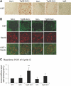
(A) Immunostaining of Ki67 in kidneys of mice. Immunohistochemistry was performed in paraffin sections of kidneys from Tg26SA/+, Tg26-SA/−, and their littermates using anti-Ki67 antibody. The representative pictures (×400) are shown here. Rabbit IgG was used as a negative control. (B) Co-localization studies. Immunofluorescent staining was performed in kidney sections of these mice for Ki67 (green) and nestin (red). Ki67 has nuclear staining and nestin follows the podocyte distribution. The bottom panel shows overlapping pictures of Ki67 and nestin (arrows indicate overlapping nucleus). (C) Real-time PCR for cyclin E. Total RNA was isolated from glomeruli of these mice using TRIzol. Real-time PCR was performed as described. The ratios of cyclin E to tubulin mRNA levels were obtained, and the folds of change to wild-type mice are shown (**P < 0.01 compared with SA/+ or Tg26-SA/− mice, n = 6).
Figure 5.
Figure 5.
(A) Immunostaining of VEGF in kidneys of mice. (A–D) Immunohistochemistry was performed in paraffin sections of kidneys from Tg26-SA/+, Tg26-SA/−, and their littermates using anti-VEGF antibody (×200). (E) VEGF staining was performed in developing kidneys (E18) (×200). (F) Rabbit IgG was used as a negative control (×200). (G) A low-power image of VEGF staining in Tg26-SA/+ mice is shown (×100). The representative pictures are shown. (B) Real-time PCR for VEGF164A. Total RNA was isolated from glomeruli of these mice using TRIzol. Real-time PCR was performed as described. The ratios of VEGF to tubulin mRNA levels were obtained and the folds of change to WT mice are shown (**P < 0.01 compared with SA/+ or Tg26-SA/− mice, n = 6). (C) Western blot for phosphor-VEGFR2. Glomerular lysates were used for Western blot analysis of phosphor-VEGFR2. Total VEGFR2, PECAM-1, and β-actin were used as the controls. Four independent experiments were performed. For each experiment, we used the glomeruli isolated from one mouse per each group. Representative blots are shown.
Figure 6.
Figure 6.
(A) Co-immunostaining was performed in kidney sections of Tg26 mice and their littermates using rabbit polyclonal anti-pStat3 and mouse monoclonal anti-nestin antibodies. The representative pictures are shown (×400). The third panel shows overlapping pictures and arrows indicate overlapping cells. (B) Western blot. Primary cultures of podocytes from both Tg26 and control Stat3 SA/+ and Stat3 SA/− mice were lysed for Western blot analysis with anti-pStat3, anti-Stat3, anti-synaptopodin, anti-podocin, anti-WT-1, anti-nestin, anti-β-actin, and anti-Nef antibodies. (C) Immunostaining. Primary cultures of podocytes isolated from Tg26-SA/+ and Tg26- SA/− mice were placed on fibronectin-coated coverslips for immunostaining with anti-synaptopodin, anti-podocin, anti-WT1, or IgG control. The representative pictures (×400) are shown; scale bars = 10 mm. The matched exposures were used for taking these pictures. A nuclear staining for synaptopodin was noticed and is likely nonspecific.
Figure 7.
Figure 7.
(A) Immunostaining of pStat3 in kidneys of mice. Immunohistochemistry was performed in kidney sections of Tg26-SA/+, Tg26-SA/−, and their littermates using rabbit polyclonal anti-pStat3 antibody (Cell Signaling). The representative pictures of tubulo-interstitial areas are shown (×400). (B) Western blot. HK2 cells were infected with a retroviral construct expression Nef as described. Infected cells were lysed for Western blot analysis for pStat3, total Stat3, and Nef. (C) Renal tubuli from both Tg26 and Stat3 SA/+ and Stat3 SA/− mice were isolated and lysed for Western blot analysis with anti-pStat3, anti-total Stat3, anti-β-actin, and anti-Nef antibodies. (D and E) Real-time PCR for ICAM1 and SOCS3. Total RNA was isolated from renal tubules of these mice using TRIzol. Real-time PCR was performed as described. The ratios of ICAM1 and SOCS3 to tubulin mRNA levels were obtained and the folds of change to WT mice are shown (**P < 0.01 compared with Tg26-SA/+ or SA/− mice, n = 4).

Comment in

References

    1. United States Renal Data System: 2005 Annual Data Report: Atlas of End-Stage Renal Disease in the United States, Minneapolis, MN, USRDS Coordinating Center, 2005 - PubMed
    1. Wyatt CM, Klotman PE: HIV-1 and HIV-associated nephropathy 25 years later. Clin J Am Soc Nephrol 2[Suppl 1]: S20–S24, 2007 - PubMed
    1. Wyatt CM, Klotman PE: HIV-associated nephropathy: A case study in race and genetics. Am J Kidney Dis 47: 1084–1085, 2006 - PubMed
    1. D'Agati V, Appel GB: HIV infection and the kidney. J Am Soc Nephrol 8: 138–152, 1997 - PubMed
    1. Bruggeman LA, Dikman S, Meng C, Quaggin SE, Coffman TM, Klotman PE: Nephropathy in human immunodeficiency virus-1 transgenic mice is due to renal transgene expression. J Clin Invest 100: 84–92, 1997 - PMC - PubMed

Publication types

Substances

LinkOut - more resources