. 2009 Sep 18;284(38):25854-66.
doi: 10.1074/jbc.M109.033472. Epub 2009 Jul 16.

Neutrophil MMP-9 proenzyme, unencumbered by TIMP-1, undergoes efficient activation in vivo and catalytically induces angiogenesis via a basic fibroblast growth factor (FGF-2)/FGFR-2 pathway

Affiliations

Neutrophil MMP-9 proenzyme, unencumbered by TIMP-1, undergoes efficient activation in vivo and catalytically induces angiogenesis via a basic fibroblast growth factor (FGF-2)/FGFR-2 pathway

Veronica C Ardi et al. J Biol Chem. .

Abstract

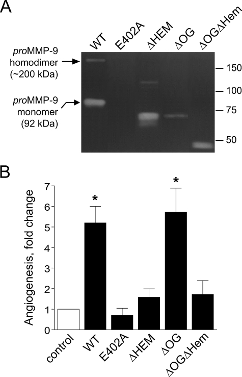
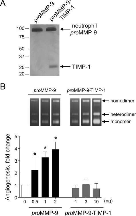
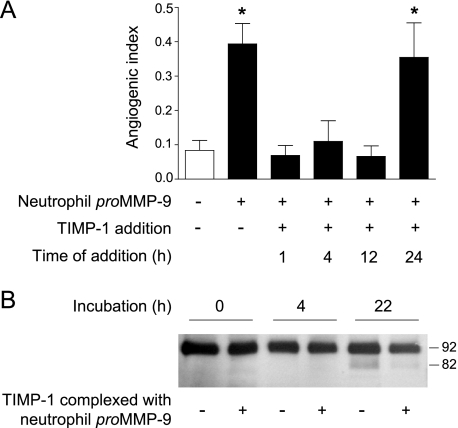
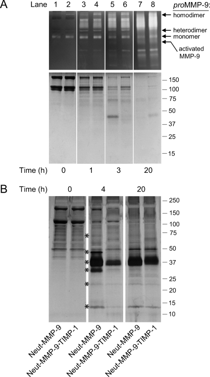
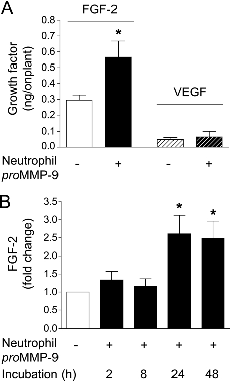
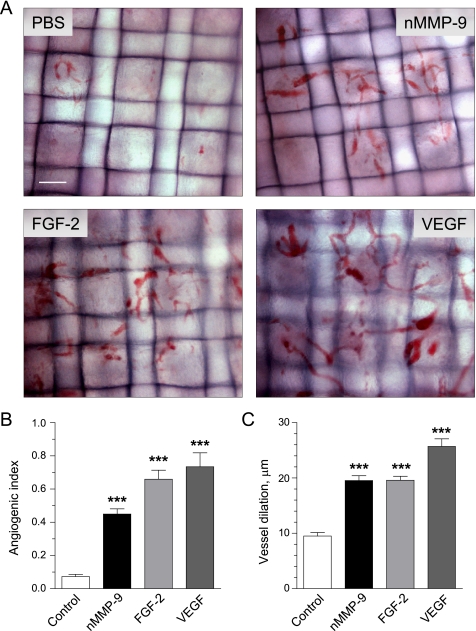
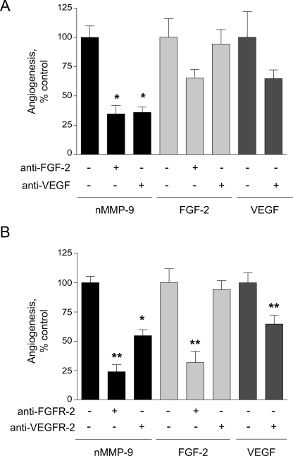
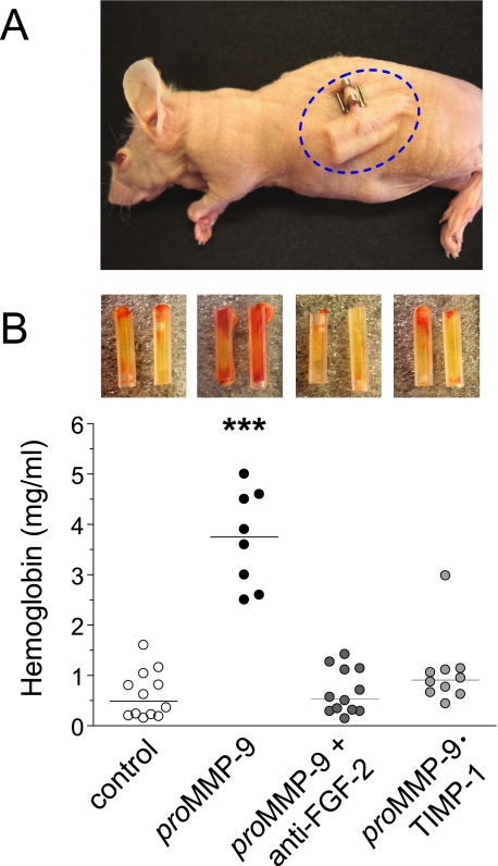
The structural and catalytic requirements for neutrophil MMP-9 proenzyme (proMMP-9) to induce angiogenesis were investigated using a quantitative angiogenesis model based on grafting of collagen onplants onto the chorioallantoic membrane of chick embryos. Both physiological activation of neutrophil proMMP-9 and proteolytic activity of the generated MMP-9 enzyme were critically dependent on the tissue inhibitor of metalloproteinase (TIMP)-free status of the zymogen. The presence of an intact active site and hemopexin domain were required for full angiogenesis-inducing activity of the MMP-9 enzyme. Timed additions of TIMP-1 to the onplants containing TIMP-free neutrophil proMMP-9 indicated that in vivo activation of the zymogen occurred during the first 24 h after grafting. Within the onplant tissue, MMP-9 activation was accompanied by proteolytic modifications of fibrillar collagen and an influx of host proteins, the rate of which depended on the TIMP-free status of the zymogen. By quantifying the levels of host angiogenic factors, we demonstrated that basic fibroblast growth factor (FGF-2) was a major cytokine becoming bioavailable in the onplant tissue undergoing a neutrophil proMMP-9-mediated angiogenic switch. Inhibition of angiogenesis with specific function-blocking antibodies further indicated an involvement of a FGF-2/FGFR-2 pathway in neutrophil proMMP-9-induced angiogenesis. The enhanced angiogenesis catalyzed by neutrophil MMP-9 appears to evoke also a localized, low threshold level vascular endothelial growth factor (VEGF)/VEGFR-2 pathway, likely functioning in the formation and/or stabilization of blood vessels. That neutrophil proMMP-9, unencumbered by TIMP-1, directly mediates FGF-2-dependent angiogenesis was also demonstrated in our quantitative mouse angiogenesis model employing subcutaneous collagen implants, thus implicating the novel TIMP-free MMP-9/FGF-2/FGFR-2 pathway in proMMP-9-induced angiogenesis in a mammalian setting.


Figures

FIGURE 1.
FIGURE 1.
Enzymatic and functional analyses of recombinant forms of human MMP-9. A, zymography analysis of MMP-9 variants. The full-length WT and catalytically inactive mutant (E402A) of MMP-9 and domain-deletion MMP-9 variants lacking the hemopexin domain (ΔHem), the linker domain containing O-linked glycans (ΔOG), and both OG and Hem domains (ΔOGΔHem) were analyzed by gelatin zymography (all at 0.02 pmol/lane). Arrows on the left indicate the positions of homodimer and monomer forms of wild type proMMP-9. Positions of the molecular mass markers in kDa are indicated on the right. B, angiogenic potential of MMP-9 variants. Individual recombinant MMP-9 proteins were added at 1–2 ng per collagen onplant (0.4–0.7 nm). The onplants were grafted on the CAM of day-10 chick embryos, and angiogenic blood vessels were scored within 72–96 h. Data are presented as -fold changes in angiogenesis (means ± S.E. from three independent experiments) over control levels in onplants supplemented with no MMP-9. *, p < 0.01.
FIGURE 2.
FIGURE 2.
Angiogenesis-inducing capacity of neutrophil proMMP-9 critically depends on TIMP-free status of the zymogen. A, silver staining of neutrophil TIMP-free proMMP-9 and proMMP-9 complexed at 1:1 molar ratio with TIMP-1. Neutrophil proMMP-9 and its stoichiometric complex with TIMP-1 were purified by gelatin-Sepharose affinity chromatography and were visualized by silver staining after SDS-PAGE under reducing conditions. Positions of protein bands corresponding to the monomer of proMMP-9 (92 kDa) and TIMP-1 (28 kDa) are indicated on the right. Positions of molecular mass markers in kDa are indicated on the left. B, angiogenic potential of the neutrophil proMMP-9 and proMMP-9·TIMP-1 complex. Purified proMMP-9 (black bars) and a stoichiometric complex of proMMP-9 with TIMP-1 (gray bars) were incorporated into collagen onplants at increasing concentrations indicated at the bottom of the graph (ng per onplant). Control onplants (open bar; 0 ng) were not supplemented with any additional exogenous factors. The levels of angiogenesis were determined at 70–76 h after grafting onplants onto the CAM of chick embryos. Data are presented as -fold changes in angiogenesis over control levels (means ± S.E. from 2 independent experiments). *, p < 0.05. Inset, zymographs depict gelatinolytic activity of MMP-9 preparations (equivalent of 1 onplant/lane). Positions of the homodimer, heterodimer, and monomer of MMP-9 are indicated on the right.
FIGURE 3.
FIGURE 3.
TIMP-free neutrophil proMMP-9 exerts angiogenic activity within first 24 h after onplant grafting. A, time-course analysis of TIMP-1-mediated inhibition of angiogenesis. Collagen onplants were supplemented with neutrophil proMMP-9 (2 ng/onplant) and grafted on the CAM. At the indicated time points (h), TIMP-1 was applied at 3× molar excess (+) over neutrophil proMMP-9 (solid bars). Control onplants contained collagen with no MMP-9 (open bar). The data are presented as the means ± S.E. of the angiogenic index determined 77 h after onplant grafting. Presented is one of two independent experiments. *, p < 0.05. B, time-course analysis of the activation of TIMP-free proMMP-9 in collagen onplants. Neutrophil proMMP-9 alone or in a stoichiometric complex with TIMP-1 (+) was incorporated into collagen onplants at 3 nm. At the indicated time periods after grafting on the CAM, the onplants were harvested, and their contents extracted with detergent lysis buffer. The MMP-9 activation in the extracts was analyzed by Western blotting with a mixture of MMP-9-specific mAbs (7-11C, 8-3H, and 6-6B) after SDS-PAGE under reducing conditions. Positions of MMP-9 zymogen (92 kDa) and activated enzyme (82 kDa) are indicated on the right.
FIGURE 4.
FIGURE 4.
Activation and proteolytic activity of neutrophil proMMP-9 and influx of host proteins into angiogenic onplants depends on TIMP-free status of the zymogen. A, time course of activation and gelatinolytic activity of neutrophil proMMP-9 and proMMP-9·TIMP-1 complex in vivo. The purified TIMP-free proMMP-9 (lanes 1, 3, 5, and 7) and proMMP-9·TIMP-1 complex (lanes 2, 4, 6, and 8) were incorporated at 3 nm into collagen onplants, which were grafted onto the CAM of chick embryos. At the indicated time intervals (h), the onplants were harvested from the CAM, extracted, processed, and analyzed by gelatin zymography (top panels) and Coomassie staining (bottom panel) of an SDS-PAGE gel run under reducing conditions. Positions of the proMMP-9 homodimer, heterodimer, and monomer and the activated MMP-9 enzyme are indicated on the right of gelatin zymographs. Positions of molecular mass markers in kDa are indicated on the right. B, influx of host proteins into collagen onplants supplemented with neutrophil proMMP-9. Collagen onplants were supplemented with TIMP-free neutrophil proMMP-9 (Neut-MMP-9) or neutrophil proMMP-9 complexed with TIMP-1 (Neut-MMP-9·TIMP-1) at 0.7 nm. At indicated time points (h) after grafting on the CAM, the onplants were harvested and extracted. The onplant extracts were separated by SDS-PAGE under non-reducing conditions, and the protein bands were visualized by silver staining. The positions of host proteins differentially influxed into the onplants containing Neut-MMP-9 versus Neut-MMP-9·TIMP-1 are indicated by asterisks. Positions of molecular weight markers (in kDa) are indicated on the right. White vertical lanes separating individual time points were placed artificially over the images of whole gels for visual purposes (A and B).
FIGURE 5.
FIGURE 5.
Increase in bioavailable FGF-2 in MMP-9-supplemented collagen onplants. A, neutrophil proMMP-9 mediates increase of bioavailable FGF-2 in angiogenic onplants. Collagen onplants were supplemented with 2 ng of neutrophil proMMP-9 (+) or buffer control only (−). After 24-h incubation on the CAM, the onplants were harvested and extracted with detergent lysis buffer. The levels of FGF-2 and VEGF were measured in the extracts by a capture ELISA. B, neutrophil proMMP-9 induces time-dependent accumulation of FGF-2. Collagen onplants with (+) or without (−) 2 ng of neutrophil proMMP-9 were harvested at the indicated time points (h) after grafting on the CAM. The onplant extracts were analyzed for the levels of FGF-2 by a specific capture ELISA. *, p < 0.01.
FIGURE 6.
FIGURE 6.
Morphological analysis of angiogenic vasculature induced by different growth factors and neutrophil proMMP-9. Angiogenesis was induced by neutrophil proMMP-9 (nMMP-9), FGF-2, and VEGF incorporated into collagen mixture at 2.5, 10, and 15 ng per onplant, respectively. Control onplants were supplemented with no growth factors. A, morphology of blood vessels in collagen onplants. Angiogenic vasculature was visualized in the plane of the upper mesh, and digital images were captured at original 100× magnification. Bar, 100 μm. B, levels of angiogenesis induced by different pro-angiogenic molecules were determined as angiogenic indices at 72 h after onplants were grafted on the CAM. Presented are cumulative data (means ± S.E.) from 4 independent experiments performed with 4–6 embryos (each carrying 5–7 onplants) per condition. C, blood vessel dilation was determined in the digital images by measuring the diameters of maximally enlarged vessels. Data are the means ± S.E. from a representative experiment. ***, p < 0.001.
FIGURE 7.
FIGURE 7.
Neutrophil proMMP-9 induces angiogenesis via FGF-2-mediated pathway. To induce angiogenesis, collagen onplants were supplemented with neutrophil proMMP-9 (nMMP-9, 2 ng) (black bars), FGF-2 (10 ng) (light gray bars), and VEGF (15 ng) (dark gray bars). Additionally, function-blocking anti-FGF-2 or anti-VEGF antibodies (A) and anti-FGFR-2 or VEGFR-2 antibodies (B) were incorporated (+) at 20 μg/ml to probe for the involvement of FGF-2/FGFR-2 and VEGF/VEGFR-2 pathways in MMP-9-induced angiogenesis. Non-immune IgGs were added in control (−/−) onplants. Inhibition of angiogenesis is presented as a percentage of control (100%) in the absence (–/–) of function-blocking antibodies determined from cumulative data from 3 to 9 independent experiments for each condition variant. Data are the means ± S.E. * and **, p < 0.05 and p < 0.01, respectively.
FIGURE 8.
FIGURE 8.
Neutrophil TIMP-free MMP-9 induces FGF-2-mediated angiogenesis in a murine angiotube model. A, immunodeficient athymic nu/nu mice were implanted subcutaneously with 4 silicon tubes (2 angiotubes per each flank) filled with collagen containing 3 nm neutrophil proMMP-9 alone or with anti-FGF-2 function-blocking antibody (50 μg/ml) or collagen supplemented with 3 nm neutrophil proMMP-9·TIMP-1 complex. B, after a 10–12-day incubation, the angiotubes were excised and photographed (insets above the corresponding scattergrams), and their hemoglobin contents were measured to determine relative levels of angiogenesis (scattergraph). ***, p < 0.0001 in comparison with all other variants.

References

    1. Vu T. H., Werb Z. (ed) (1998) in Matrix Metalloproteinases ( Parks W. C., Mecham R. P. eds) pp. 115–148, Academic Press, San Diego
    1. Opdenakker G., Van den Steen P. E., Dubois B., Nelissen I., Van Coillie E., Masure S., Proost P., Van Damme J. (2001) J. Leukocyte Biol. 69, 851–859 - PubMed
    1. Van den Steen P. E., Dubois B., Nelissen I., Rudd P. M., Dwek R. A., Opdenakker G. (2002) Crit. Rev. Biochem. Mol. Biol. 37, 375–536 - PubMed
    1. Parks W. C., Wilson C. L., López-Boado Y. S. (2004) Nat. Rev. Immunol. 4, 617–629 - PubMed
    1. Björklund M., Koivunen E. (2005) Biochim. Biophys. Acta 1755, 37–69 - PubMed

Publication types

MeSH terms

Substances