. 2009 Aug 21;325(5943):1010-4.
doi: 10.1126/science.1176088. Epub 2009 Jul 16.

Structure and mechanism of a Na+-independent amino acid transporter

Affiliations

Structure and mechanism of a Na+-independent amino acid transporter

Paul L Shaffer et al. Science. .

Abstract

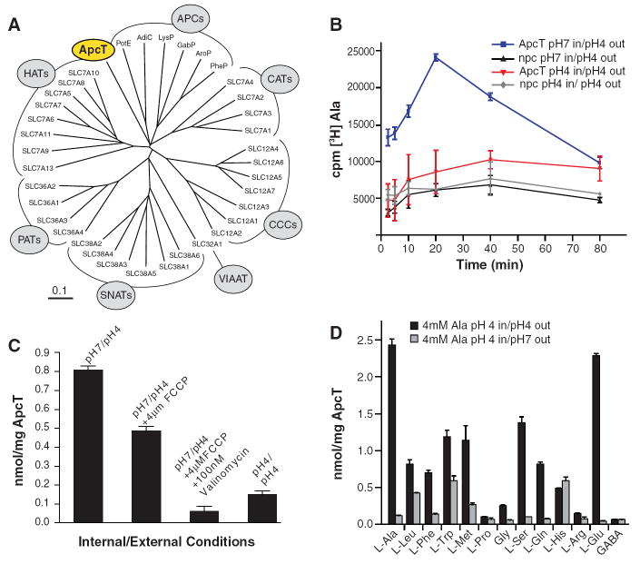
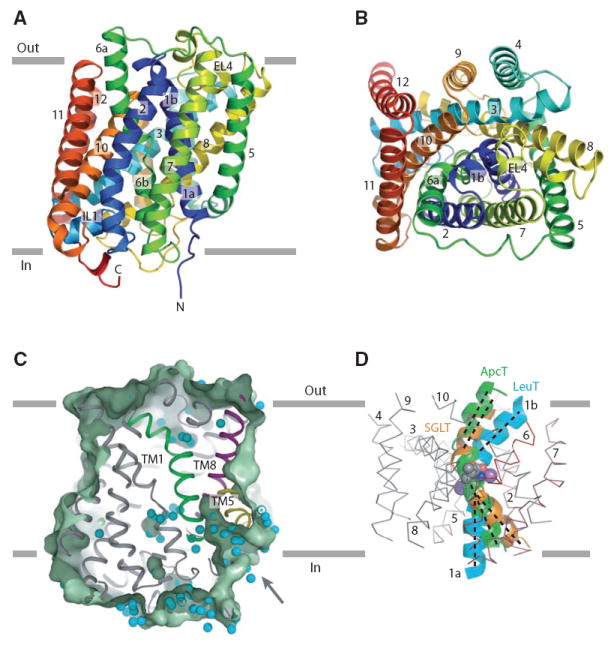
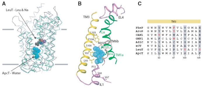
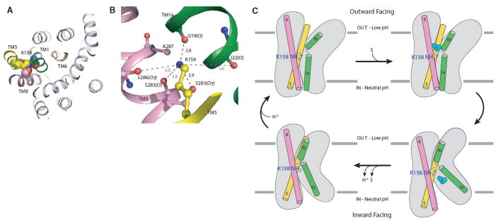
Amino acid, polyamine, and organocation (APC) transporters are secondary transporters that play essential roles in nutrient uptake, neurotransmitter recycling, ionic homeostasis, and regulation of cell volume. Here, we present the crystal structure of apo-ApcT, a proton-coupled broad-specificity amino acid transporter, at 2.35 angstrom resolution. The structure contains 12 transmembrane helices, with the first 10 consisting of an inverted structural repeat of 5 transmembrane helices like the leucine transporter LeuT. The ApcT structure reveals an inward-facing, apo state and an amine moiety of lysine-158 located in a position equivalent to the sodium ion site Na2 of LeuT. We propose that lysine-158 is central to proton-coupled transport and that the amine group serves the same functional role as the Na2 ion in LeuT, thus demonstrating common principles among proton- and sodium-coupled transporters.


Figures

Fig. 1
Fig. 1. ApcT is a broad specificity amino acid transporter
(A) Dendrogram of human and selected bacterial APC transporters. Branches corresponding to prokaryotic APCs, CATs, CCCs, VIATT, SNATs, PATs and HATs are defined in table S1. (B) [3H]-Ala uptake is pH dependent. ApcT and no protein control proteoliposomes were loaded with buffer at pH 7 (blue and black, respectively) or pH 4 (red and grey, respectively). Uptake experiment was performed by diluting these liposomes in a pH 4 buffer supplemented with 500nM [3H]Ala. Error bars represent SEM of triplicate measurements. (C) Effect of FCCP and valinomycin treatment on [3H]-Ala uptake. Proteoliposomes were loaded with 100mM KCl, and either pH7 or pH4 20mM citrate buffer. Uptake experiment was performed at 30°C in 20mM citrate buffer pH4, 100mM KCl, 750nM L-[3H] Ala and in the presence or absence of 4μM FCCP and/or 100nM valinomycin. Time points were taken at 20 min. (D) Ala and Glu are the preferred substrates of ApcT in counterflow experiments. Proteoliposomes were loaded with 4mM Ala, pH 4 and uptake of the particular [3H] amino acid, at 500 nM concentration, was measured. Estimates of uptake together with non specific transport or binding are defined by the experiments at pH4 (external; black bars) whereas estimates of non specific transport or binding are defined by experiments at pH 7 (external; gray bars).
Fig. 2
Fig. 2. Architecture of ApcT
(A) Ribbon diagram of the ApcT structure, viewed parallel to the membrane, along the pseudo 2- fold axis of molecular symmetry. (B) ‘Top’ down view of ApcT from the outside. (C) Slice through a solvent accessible surface of ApcT showing a solvent accessible pathway reaching deep into the transporter. Water molecules are shown as cyan spheres. (D) Superposition of the scaffold helices TMs 3-5 and 8-10 of ApcT and vSGLT on to the equivalent elements of LeuT shows that in ApcT, TM1b is closed to the outside and TM1a is partially open to the inside. TMs 2-10 of LeuT are shown as an α-carbon trace.
Fig. 3
Fig. 3. Substrate pocket
(A) Structural superposition of TMs 1-10 of LeuT (grey) and ApcT (green). TMs 1-10 of LeuT were superposed on that of ApcT using the DaliLite program and their Cα traces are shown. The substrate leucine and two sodium ions in LeuT are shown as spheres. C, N, O and Na atoms are grey, blue, red and violet respectively. The water molecules buried in the middle of the TM regions in ApcT are shown in cyan. (B) A closeup view of the putative solvent pocket in ApcT. TM helices 1, 3 and 6 and the IL1 and EL4 loops surrounding the solvent pocket are shown. Key residues predicted to be involved in substrate binding are shown as ball and stick models. (C) Role of TM3 in substrate binding. The sequence alignment shown is a composite of independent alignments generated by PROMALS 3D of the prokaryotic transporters (PheP, AroP, CAN1, GNP1, AdiC, LeuT and ApcT) and of ApcT alone with xCT orthologues. Residues implicated in substrate binding are highlighted in red and equivalent residues in other orthologs are highlighted by grey shading.
Fig. 4
Fig. 4. Lys158 is essential to ApcT transport function
(A) View of Lys158 bound between TMs1 and 8. (B) The amine of Lys158 forms hydrogen bonds with the carbonyl and hydroxyl oxygens of Gly19 and S283, respectively. (C) We propose that ApcT adopts an inward facing, occluded conformation when the amine group of K158 is neutral (bottom-left panel). Acidic pH promotes protonation of K158 and isomerization to an outward facing conformation (top-left). Upon substrate binding (top-right), ApcT isomerizes to an open-to-in state (bottom-right). The release of substrate and proton to the cytoplasm precedes formation of the inward facing yet occluded conformation observed in the present crystal structure (bottom-left panel).

References

    1. Jack DL, Paulsen IT, Saier MHJ. Microbiology. 2000;146:1797. - PubMed
    1. Hediger MA, et al. Pflugers Arch. 2004;447:465. - PubMed
    1. Sato H, Tamba M, Ishii T, Bannai S. J Biol Chem. 1999;274:11455. - PubMed
    1. Bannai S, Kitamuri E. J Biol Chem. 1980;255:2372. - PubMed
    1. Zimmermann N, et al. J Clin Invest. 2003;111:1815. - PMC - PubMed

Publication types

MeSH terms