. 2009 Aug 21;325(5943):1006-10.
doi: 10.1126/science.1175870. Epub 2009 Jul 16.

Bcl6 and Blimp-1 are reciprocal and antagonistic regulators of T follicular helper cell differentiation

Affiliations

Bcl6 and Blimp-1 are reciprocal and antagonistic regulators of T follicular helper cell differentiation

Robert J Johnston et al. Science. .

Abstract

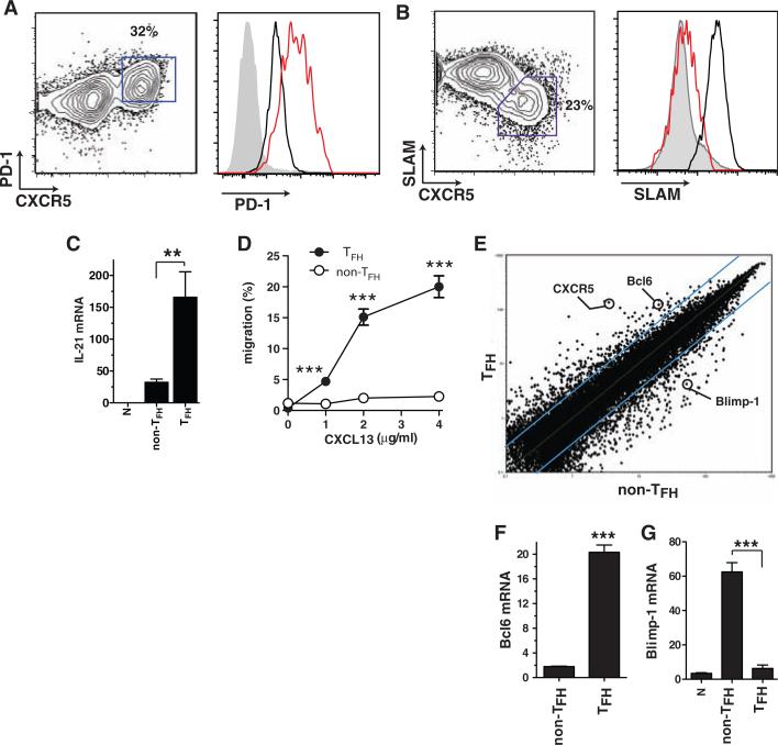
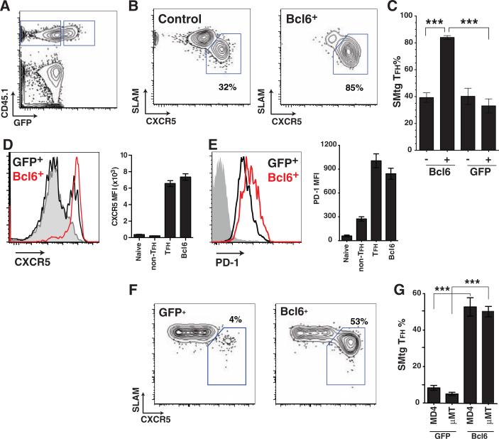
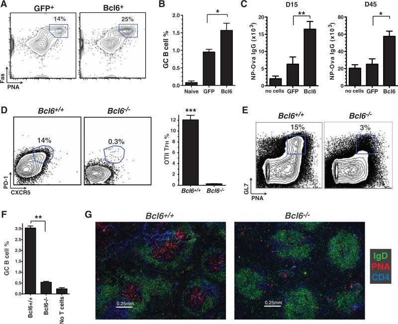
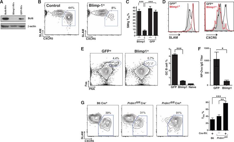
Effective B cell-mediated immunity and antibody responses often require help from CD4+ T cells. It is thought that a distinct CD4+ effector T cell subset, called T follicular helper cells (T(FH)), provides this help; however, the molecular requirements for T(FH) differentiation are unknown. We found that expression of the transcription factor Bcl6 in CD4+ T cells is both necessary and sufficient for in vivo T(FH) differentiation and T cell help to B cells in mice. In contrast, the transcription factor Blimp-1, an antagonist of Bcl6, inhibits T(FH) differentiation and help, thereby preventing B cell germinal center and antibody responses. These findings demonstrate that T(FH) cells are required for proper B cell responses in vivo and that Bcl6 and Blimp-1 play central but opposing roles in T(FH) differentiation.


Figures

Fig. 1
Fig. 1
Bcl6 is a TFH-specific transcription factor. Naïve SMtg CD4+ T cells were transferred into B6 mice. In all panels, splenocytes were analyzed 8 days after infection with LCMV. (A and B) SMtg expression of CXCR5 and PD-1 (A) or SLAM (CD150) (B). SMtg+ (CD45.1+) CD4+ gated cells are shown. CXCR5high TFH cells are boxed in fluorescence-activated cell sorter (FACS) plots. Histogram overlays depict TFH cells (red) as well as naïve CD4+ T cells (gray) and CXCR5low non-TFH SMtg cells (black). Data are representative of more than 10 independent experiments. (C) IL-21 mRNA in SMtg CD4+ T cells, normalized to the β-actin mRNA level (×10–4). **P = 0.008. (D) In vitro chemotaxis toward CXCL13 (BLC) by ex vivo SMtg CD4+ T cells. Results are expressed as percentages of SLAMlow TFH SMtg (solid circles) and SLAMhigh non-TFH SMtg (open circles) that migrated in a transwell assay. ***P ≤ 0.001. 1 μg, P = 0.001; 2 μg, P = 0.0006; 4 μg, P = 0.0006. Data are representative of three independent experiments; n = 2 per group. (E) Scatterplot of the average signal of biological replicates of TFH versus non-TFH SMtg gene expression microarray data. Blue lines indicate changes in gene expression by a factor of 3; 386 gene probes exhibited a factor of >3.0 increase in TFH. Data from one of two independent experiments are shown; n = 2 per group. (F and G) Quantitative reverse transcription PCR of Bcl6 (F) and Blimp-1 (G) mRNA expression, normalized to β-actin (×10–4). ***P < 0.0001. Data are representative of four independent experiments; n = 2 per group. Error bars in all graphs are SEM.
Fig. 2
Fig. 2
Bcl6 expression is sufficient for TFH differentiation in vivo. (A to E) Naïve SMtg CD4+ T cells were transduced with Bcl6-RV (Bcl6+) or left untransduced (control) and transferred into B6 mice subsequently infected with LCMV. (A) Gating of CD45.1+ untransduced SMtg (GFP) and Bcl6-RV+ SMtg (GFP+) in the same host. CD4+ B220 gate is shown. (B) TFH (SLAMlow CXCR5high, boxed) and non-TFH (SLAMhigh CXCR5low) differentiation of untransduced SMtg (left) and Bcl6-RV+ SMtg (right). (C) Quantitation of SMtg TFH differentiation. Mice received Bcl6-RV+ SMtg and untransduced SMtg, or GFP-RV+ and untransduced SMtg. “–,” untransduced; “+,” transduced with indicated RV. ***P < 0.0001. Data are representative of three independent experiments; n = 4 per group. (D and E) CXCR5 expression (D) and PD-1 expression (E) on naïve CD4+ T cells (gray), GFP-RV+ SMtg (black), and Bcl6-RV+ SMtg (red). Bar graphs show mean fluorescence intensity (MFI) of naïve CD4+ T cells, GFP-RV+ SMtg non-TFH, GFP-RV+ SMtg TFH, and Bcl6-RV+ SMtg. For PD-1 MFI, non-TFH versus TFH or Bcl6-RV+, P < 0.05; TFH versus Bcl6-RV+, P > 0.05. Data are representative of three independent experiments; n = 4 to 6 per group. (F and G) GFP-RV+ or Bcl6-RV+ SMtg cells were adoptively transferred separately into B cell–deficient mice (μMT) or HEL-specific BCR transgenic mice (MD4) on a μMT background. Host mice were subsequently infected with LCMV. Each group is a composite of three experiments; n = 2 (GFP-RV+ μMT), 6 (Bcl6-RV+ MD4), 6 (Bcl6-RV+ μMT), or 8 (GFP-RV+ MD4) per group. (F) Differentiation of SMtg CD4+ T cells in MD4 BCR transgenic mice. TFH cells (SLAMlow CXCR5high) are boxed. CD4+ B220 CD45.1+ GFP+ gate is shown. (G) Quantitation of SMtg TFH differentiation. GFP-RV+ versus Bcl6-RV+ in MD4, ***P < 0.0001. GFP-RV+ versus Bcl6-RV+ in μMT, ***P < 0.0001.
Fig. 3
Fig. 3
Bcl6 expression is necessary for inducing TFH B cell help in vivo. (A) Germinal center B cells (PNA+ Fas+, gated) in mice that received GFP-RV+ or Bcl6-RV+ SMtg CD4+ T cells and were subsequently infected with LCMV, analyzed at day 8 and gated on activated B cells (B220+ IgDlow). (B) Frequency of germinal center B cells of total splenocytes; n = 4 per group. Data are representative of three independent experiments. *P = 0.029. (C) GFP-RV+ or Bcl6-RV+ OT-II CD4+ T cells were transferred into B6 mice subsequently immunized with NP-Ova in alum. Control mice were immunized but received no OT-II cells. NP-Ova enzyme-linked immunosorbent assay (ELISA) was performed at day 15 and day 45; n = 6 per group. Data are representative of two independent experiments. Day 15 endpoint ELISA titers, **P = 0.008; day 45 endpoint ELISA titers, *P = 0.017.(D) Bcl6+/+ or Bcl6–/– OT-II CD4+ Tcells were transferred into congenically mismatched B6 mice subsequently immunized with Ova in alum. Splenocytes were analyzed 6 days after immunization; n = 4 per group. Data are representative of four independent experiments. OT-II+ CD44high gate is shown. Quantitation of OT-II TFH differentiation is also shown. ***P < 0.0001. (E to G) Bcl6+/+ or Bcl6–/– OT-II CD4+ T cells were cotransferred with B1-8 B cells into Icos–/– mice subsequently immunized with NP-Ova in alum; n = 2 per group. Data are representative of two independent experiments. (E) Germinal center B cells (PNA+ GL7+, boxed) 7 days after immunization. TCRβ IgDlow gate is shown. (F) Quantitation of GC B cells as percent of spleen. **P = 0.0015. (G) Germinal center histology. Spleen sections were stained with IgD (green), PNA (red), and CD4 (blue).
Fig. 4
Fig. 4
Blimp-1 and Bcl6 are antagonistic and reciprocal regulators of TFH differentiation. (A) Immunoblot of Bcl6 protein expression (and β-actin control) in transduced SMtg CD4+ T cells in vivo. (B) TFH (SLAMlow CXCR5high, boxed) differentiation of untransduced SMtg (left, “Control”) and Blimp1-RV+ SMtg (right, “Blimp-1+”) cells within a common host 8 days after LCMV infection. Gating is shown in fig. S10B. (C) Quantitation of SMtg TFH differentiation. “–,” untransduced; “+,” transduced with the indicated RV. ***P < 0.0001; n = 4 per group. Data are representative of two independent experiments. (D) SLAM and CXCR5 expression by naïve CD4+ T cells (gray), GFP-RV+ SMtg (black), and Blimp1-RV+ SMtg (red). (E and F) GFP-RV+ or Blimp1-RV+ OT-II CD4+ T cells were transferred into SAP-deficient mice subsequently immunized with NP-Ova in alum; n = 4 per group. Data are representative of three independent experiments. (E) Germinal center B cells (PNA+ Fas+, gated) in mice that received GFP-RV+ or Blimp1-RV+ OT-II CD4+ T cells. B220+ IgDlow gate is shown. Quantitation of germinal center B cells in the spleen is also shown. ***P < 0.0001. (F) NP-Ova IgG ELISA endpoint titers at day 10. *P = 0.016. (G) Purified naïve B6 and prdm1fl/fl CD45.2+ CD4+ T cells were transduced with SMtg-RV, with or without Cre-RV (Cre+ or Cre), sorted, and transferred into CD45.1+ mice subsequently infected with LCMV. FACS plots depict TFH (CXCR5high SLAMlow, boxed) differentiation of control Cre+ SMtg+ B6 cells (left), Blimp-1–sufficient Cre SMtg+ prdm1fl/fl cells (center), and Blimp-1–deficient Cre+ SMtg+ prdm1fl/fl cells (right). CD4+ CD45.1 CD44high 7AAD gate is shown. Quantitation of TFH differentiation is also shown. Data are representative of two independent experiments. **P = 0.002, ***P = 0.0006; n = 4 to 5 per group.

Comment in

References

    1. Zhu J, Paul WE. Blood. 2008;112:1557. - PMC - PubMed
    1. King C, Tangye S, Mackay C. Annu. Rev. Immunol. 2008;26:741. - PubMed
    1. Vinuesa CG, et al. Nature. 2005;435:452. - PubMed
    1. Chtanova T, et al. J. Immunol. 2004;173:68. - PubMed
    1. Rasheed AU, Rahn HP, Sallusto F, Lipp M, Müller G. Eur. J. Immunol. 2006;36:1892. - PubMed

Publication types

MeSH terms

Associated data