. 2009 Aug 21;325(5943):995-8.
doi: 10.1126/science.1173275. Epub 2009 Jul 16.

An expressed fgf4 retrogene is associated with breed-defining chondrodysplasia in domestic dogs

Affiliations

An expressed fgf4 retrogene is associated with breed-defining chondrodysplasia in domestic dogs

Heidi G Parker et al. Science. .

Abstract

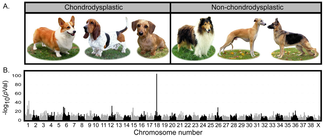
Retrotransposition of processed mRNAs is a common source of novel sequence acquired during the evolution of genomes. Although the vast majority of retroposed gene copies, or retrogenes, rapidly accumulate debilitating mutations that disrupt the reading frame, a small percentage become new genes that encode functional proteins. By using a multibreed association analysis in the domestic dog, we demonstrate that expression of a recently acquired retrogene encoding fibroblast growth factor 4 (fgf4) is strongly associated with chondrodysplasia, a short-legged phenotype that defines at least 19 dog breeds including dachshund, corgi, and basset hound. These results illustrate the important role of a single evolutionary event in constraining and directing phenotypic diversity in the domestic dog.


Figures

Figure 1
Figure 1
Results of a whole genome association analysis for chondrodysplasia across 72 breeds of dog. A). Examples of breeds used as cases (Pembroke Welsh corgi, basset hound, and dachshund pictured) and controls (collie, whippet, and German shepherd dog) in this analysis. B) Alternating shades of gray and black designate the chromosomal boundaries. The two highest peaks are found on chromosome 18 at bases 23,298,242 and 23,729,786 in CanFam2 assembly (www.genome.ucsc.edu). The peaks are less than .5 Mb apart and appear merged in the graph. Dog photos courtesy of Mary Bloom ©AKC.
Figure 2
Figure 2
Observed heterozygosity in chondrodysplastic (red) and non-chondrodysplastic (black) breeds within the associated region on chromosome 18. A) Graph of observed heterozygosity (Ho) across a 34 kb region on CFA18. Each point is the average Ho at one marker across all individuals within the group. The X axis shows the position on chromosome 18. The lines, red for chondrodysplastic and black for non-chondrodysplastic, show the trend in heterozygosity across the region by LOWESS (locally weighted least squares) best fit to the data. The average Ho for controls across the 24 kb homozygous region is 0.10. B) Schematic of the region that is homozygous and identical in chondrodysplastic breeds. Gene 1 is a pseudogene similar to thiorodoxin domain containing 1 (txndc1). Gene 2 is the 3’ end of semaphorin 3c (sema3c). The green boxes labeled putative regulatory regions are conserved in both sequence and context in all mammals for which genome data are available. A five kb insertion (red rectangle) was found within the fourth LINE between the two putative regulatory elements. The insertion contains an fgf4 retrogene. Arrangement of genes and conserved regions are per the CanFam2 assembly (www.genome.ucsc.edu).
Figure 3
Figure 3
Comparison of insert to source FGF4 gene. The first row on the figure displays the alignment of the insert sequence to the source FGF4 sequence. FGF4 has three coding exons represented by the green boxes on the graph and begins at CFA18 position 51439420 and ends at position 51441146. All three exons are present in the insert which aligns between positions 51439178 and 51442902. Tthe insert includes 242 bases upstream of the start site and 1756 bases downstream of the stop codon followed by a polyA repeat. A 13 base sequence (AAGTCAGACAGAG) derived from the insert site, indicated by a blue R on the figure, is repeated at both ends of the insert. The second line shows the coding sequence of FGF4 with the size of the exons and introns labeled. Alignment of the mouse promoter and enhancer sequences are indicated by the blue lines directly above the dog/human/mouse/rat conservation track shown at the bottom of the figure (www.genome.ucsc.edu; TRED: http://rulai.cshl.edu/cgi-bin/TRED/tred.cgi?process=home)(34). Coding sequence is predicted based on sequence similarity of translated proteins (accession # XM_540801).
Figure 4
Figure 4
Restriction fragment length polymorphism genotyping of FGF4, the fgf4 retrogene, and the fgf4 transcript from chondrodysplastic dogs. A) A 505 bp fragment was amplified from gel extracted PCR products containing the fgf4 retrogene, the source FGF4 3’UTR, and from messenger cDNA created from articular cartilage of the distal humerus (lanes 1–2) and the proximal humerus (lanes 3–4) of a 4 week old chondrodysplastic dog. Each fragment was cut to completion with restriction enzyme BsrB1 and run on a 2% agarose gel. The cDNA shows alleles specific to both the source gene and the retrogene verifying expression of the latter. B) The same experiment was done on non-chondrodysplastic fetal dogs (a spaniel mix in lanes 5–6, hound mix in lanes 7–8). Lanes 5 and 7 are amplified from the cDNA from proximal tibia. Lanes 6 and 8 are from cDNA from distal femur. Genotypes from the source gene and cDNA are identical as no other copy of FGF4 is present. C) Genes were amplified in cDNA from articular cartilage from the proximal humerus in an adult chondrodysplastic (Shih Tzu) and non-chondrodysplastic (Siberian Husky) dog. Though RNA levels were low in these tissues, expression of CD36 and Sema3C were strong, but neither the source FGF4 nor the fgf4 retrogene could be detected.

Comment in

  • Genetics. More than just a copy.
    Kaessmann H. Kaessmann H. Science. 2009 Aug 21;325(5943):958-9. doi: 10.1126/science.1178487. Science. 2009. PMID: 19696341 No abstract available.

References

    1. Wayne RK, Ostrander EA. Trends Genet. 2007 Nov;23:557. - PubMed
    1. Fondon JW, 3rd, Garner HR. Proc Natl Acad Sci U S A. 2004 Dec 28;101:18058. - PMC - PubMed
    1. Kirkness EF, et al. Science. 2003;301:1898. - PubMed
    1. Cruz F, Vila C, Webster MT. Mol Biol Evol. 2008 Nov;25:2331. - PubMed
    1. Stockard CR. The Genetic and Endocrinic Basis for Differences in Form and Behavior. Philadelphia: The Wistar Institute of Anatomy and Biology; 1941.

Publication types

MeSH terms