. 2009 Jul 17;325(5938):321-5.
doi: 10.1126/science.1173755.

The human SepSecS-tRNASec complex reveals the mechanism of selenocysteine formation

Affiliations

The human SepSecS-tRNASec complex reveals the mechanism of selenocysteine formation

Sotiria Palioura et al. Science. .

Abstract

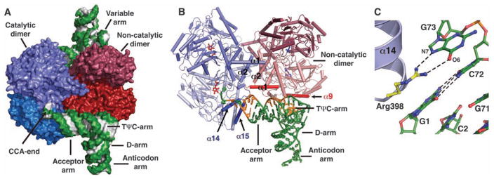
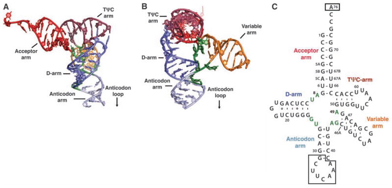
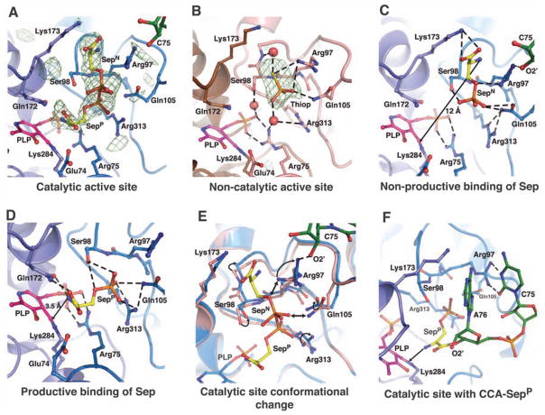
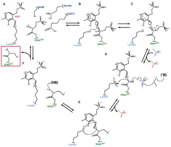
Selenocysteine is the only genetically encoded amino acid in humans whose biosynthesis occurs on its cognate transfer RNA (tRNA). O-Phosphoseryl-tRNA:selenocysteinyl-tRNA synthase (SepSecS) catalyzes the final step of selenocysteine formation by a poorly understood tRNA-dependent mechanism. The crystal structure of human tRNA(Sec) in complex with SepSecS, phosphoserine, and thiophosphate, together with in vivo and in vitro enzyme assays, supports a pyridoxal phosphate-dependent mechanism of Sec-tRNA(Sec) formation. Two tRNA(Sec) molecules, with a fold distinct from other canonical tRNAs, bind to each SepSecS tetramer through their 13-base pair acceptor-TPsiC arm (where Psi indicates pseudouridine). The tRNA binding is likely to induce a conformational change in the enzyme's active site that allows a phosphoserine covalently attached to tRNA(Sec), but not free phosphoserine, to be oriented properly for the reaction to occur.


Figures

Fig. 1
Fig. 1
Structure of human SepSecS in complex with unacylated tRNASec. (A) Surface representation of the physiological complex of SepSecS with tRNASec. The subunits of the catalytic dimer are dark and light blue, those of the noncatalytic dimer are dark and light red; the backbone and the bases of tRNASec are green and gray, respectively. (B) The catalytic dimer interacts with the acceptor arm of tRNASec through helices α14 and α15 (blue). The α1 helix (red) of the noncatalytic dimer interacts with the rest of the acceptor-TΨC arm. The regions of tRNASec that interact with SepSecS are shown in orange; the rest is green. One tRNASec molecule is shown for clarity. (C) Interactions between the discriminator base G73 and the conserved Arg398 in the α14-β11 loop. The protein side chains are gold, and tRNASec is green.
Fig. 2
Fig. 2
Structure of human tRNASec. (A) Ribbon diagram of the human tRNASec molecule observed in complex with SepSecS. The major structural elements are colored as follows: the acceptor arm is red, the D-arm is blue, the anticodon arm is light blue, the variable arm is orange, and the TΨC arm is dark red. (B) The view is rotated by ~90° clockwise around the vertical axis. (C) Secondary structure diagram of human tRNASec derived from the crystal structure.
Fig. 3
Fig. 3
Ligand binding to the active sites in the SepSecS-tRNASec complex. In (A), (C), (D), (E), and (F), the catalytic PLP-monomer is purple, the P-loop monomer is light blue, Sep is gold, tRNA is green, and PLP is magenta. In (A) and (B), the unbiased omit electron density map (green mesh) is contoured at 3.5 σ. (A) Phosphoserine binds to the catalytic sites in two orientations (SepP and SepN). (B) Thiop (orange) binds only to the non-catalytic site. The PLP-monomer is brown, and the P-loop monomer is pink. (C) The SepN amino group is ~12 Å away from the Schiff base. Arg97, Gln105, and Arg313 coordinate phosphate, and the carboxyl group interacts with Lys173. (D) The amino group of SepP interacts with Gln172 and is 3.5 Å away from the Schiff base. The carboxyl group interacts with Gln172, whereas Ser98, Gln105, and Arg313 coordinate phosphate. (E) The P-loop adopts different conformation after tRNASec binding. The noncatalytic dimer is pink, the catalytic dimer is light blue. Steric clashes between the noncatalytic P-loop and SepN are shown (double arrow). (F) A model of CCA-SepP (gold) in the catalytic active site. A76 binds between the side chains of Arg97 and Lys173, whereas C75 interacts with Arg97.
Fig. 4
Fig. 4
The PLP-dependent mechanism of Sep to Sec conversion. (A) The phosphoseryl moiety of Sep-tRNASec is bound to the active site similar to SepP. The amino group is oriented for attack on the Schiff base, whereas the phosphoryl group is stabilized by the side chains of Ser98, Gln105, and Arg313. Hydrogen bonds are shown in dashed lines. (B) After the formation of the external aldimine, the side chain of Lys284 rearranges and abstracts the Cα proton from Sep. The protonated pyridine ring of PLP stabilizes the carbanion. (C) Electron delocalization leads to a rapid β-elimination of phosphate and to the formation of dehydroalanyl-tRNASec. Free phosphate dissociates, and selenophosphate binds to the active site. (D) An unidentified base (B) activates water that hydrolyzes selenophosphate. Free phosphate dissociates again, and selenium attacks the dehydroalanyl-tRNASec. Lys284 returns the proton to the Cα carbon, and the selenocysteinyl moiety is formed. (E) The reaction of reverse transaldimination is shown. Lys284 forms the Schiff base, with PLP leading to a release of the oxidized form of Sec-tRNASec (red box). (F) The free amino group of Sec-tRNASec is protonated, and the active site of SepSecS is regenerated.

References

    1. Böck A, Thanbichler M, Rother M, Resch A. In: Aminoacyl-tRNA Synthetases. Ibba M, Francklyn C, Cusack S, editors. Landes Bioscience; Georgetown, TX: 2005. pp. 320–327.
    1. Su D, et al. IUBMB Life. 2009;61:35. - PubMed
    1. Ambrogelly A, Palioura S, Söll D. Nat Chem Biol. 2007;3:29. - PubMed
    1. Sheppard K, et al. Nucleic Acids Res. 2008;36:1813. - PMC - PubMed
    1. Bösl MR, Takaku K, Oshima M, Nishimura S, Taketo MM. Proc Natl Acad Sci USA. 1997;94:5531. - PMC - PubMed

Publication types

MeSH terms

Associated data

LinkOut - more resources